

Review
doi: 10.1128/IAI.01369-10.
Epub 2011 Feb 22.
How to become a top model: impact of animal experimentation on human Salmonella disease research
Affiliations
- PMID: 21343352
- PMCID: PMC3088149
- DOI: 10.1128/IAI.01369-10
Item in Clipboard
Review
How to become a top model: impact of animal experimentation on human Salmonella disease research
Infect Immun.
2011 May.
Abstract
Salmonella serotypes are a major cause of human morbidity and mortality worldwide. Over the past decades, a series of animal models have been developed to advance vaccine development, provide insights into immunity to infection, and study the pathogenesis of human Salmonella disease. The successive introduction of new animal models, each suited to interrogate previously neglected aspects of Salmonella disease, has ushered in important conceptual advances that continue to have a strong and sustained influence on the ideas driving research on Salmonella serotypes. This article reviews important milestones in the use of animal models to study human Salmonella disease and identify research needs to guide future work.
Figures
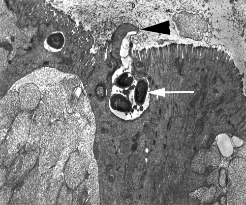
S. Typhimurium invades the intestinal epithelium. A transmission electron micrograph of the ileal mucosa of a calf experimentally infected with S. Typhimurium is shown. Local degeneration of microvilli and formation of membrane extrusions at the apical surface (arrowhead) of an enterocyte are associated with internalization of bacteria (arrow). Akio Takeuchi recorded similar images in his pioneering studies of epithelial invasion by S. Typhimurium in the guinea pig (84).
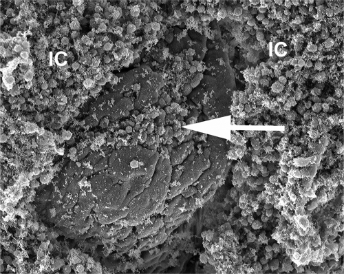
Neutrophils migrate into the intestinal lumen in response to S. Typhimurium infection. A scanning electron micrograph of the ileal mucosa of a calf experimentally infected with S. Typhimurium is shown. The image shows epithelial erosion at the tip of an intestinal villus (arrow) and marked accumulation of inflammatory cells (IC) on the luminal surface of the intestinal mucosa.
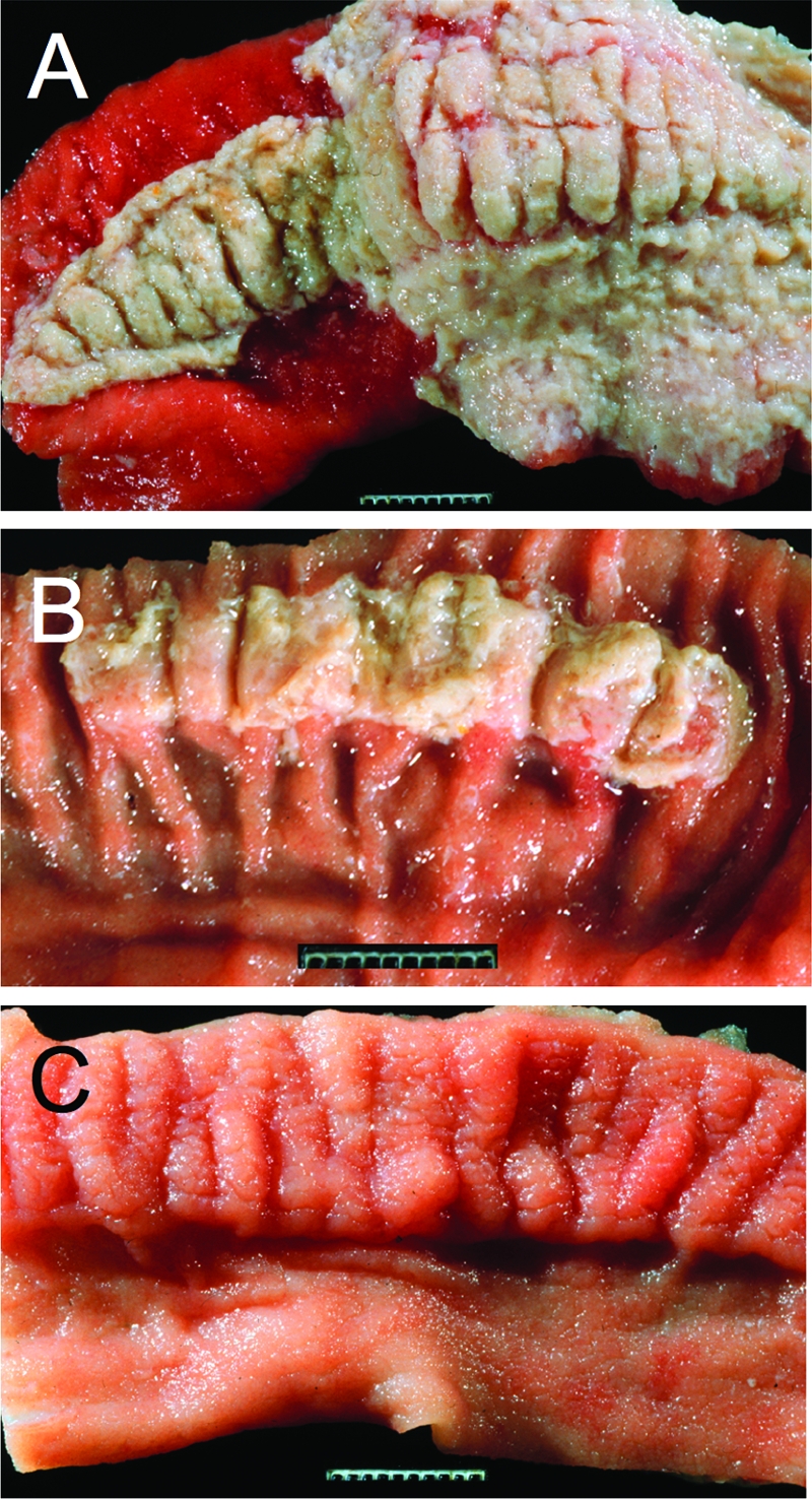
T3SS-1 and T3SS-2 contribute to intestinal inflammation in the calf gastroenteritis model. (A to C) Gross pathological appearance of the luminal surface of the terminal ileum collected 2 days after oral infection of calves with the S. Typhimurium wild-type (A), a T3SS-2-deficient mutant (B), or a T3SS-1-deficient mutant (C). Compared to the severe lesions caused by infection with the wild type (severe acute fibrinopurulent necrotizing enteritis with segmental or continuous pseudomembrane formation [A]), intestinal inflammation is reduced in calves infected with a T3SS-2-deficient mutant (moderate to marked subacute fibrinopurulent enteritis often confined to Peyer's patches [B]) and absent in calves infected with a T3SS-1-deficient mutant (normal Peyer's patch and ileal mucosa [C]). (Reprinted from reference .)
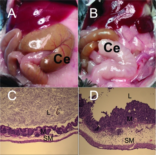
Inflammation of the cecum in the mouse colitis model. (A) Normal appearance of the cecum (Ce) in a mock-infected control animal. (B) Shrunken and edematous cecum (Ce) from a S. Typhimurium-infected mouse. (C and D) Histopathological appearance of the murine cecum of a mock-infected mouse (C) or a mouse infected with the S. Typhimurium wild type (D) 72 h after infection. Note that S. Typhimurium infection (D) is associated with severe diffuse neutrophil infiltrate in the mucosa (M) and submucosa (SM) and severe edema in the submucosa. L, lumen. (The images in panels C and D were reprinted from reference .)
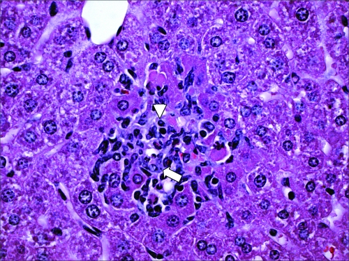
S. Typhimurium disseminates systemically in the mouse colitis model. A microgranuloma in the liver of a streptomycin-pretreated mouse infected with S. Typhimurium is shown. The image shows focal accumulation of lymphocytes (arrowhead) and epithelioid macrophages (arrow) in a section of the liver.
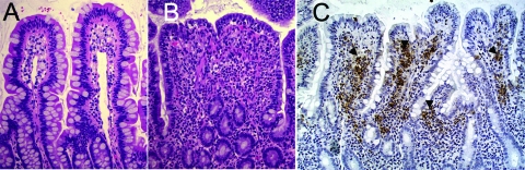
The rhesus macaque (Macaca mulatta) model. (A) Image of the uninfected ileal mucosa. (B) Section of the intestinal mucosa 8 h after infection of a ligated ileal loop with S. Typhimurium. Note the blunting of intestinal villi and the marked infiltration of inflammatory cells, predominately neutrophils. (C) Immunohistochemical labeling (brown precipitate) of S. Typhimurium in the intestinal mucosa of a rhesus macaque (Macaca mulatta) 8 h after inoculation of a ligated ileal loop with S. Typhimurium. Note the marked immunolabeling (arrowheads) of S. Typhimurium in the lamina propria, which illustrates the invasive nature of the infection.
References
-
- Arthur G., et al. 2001. Trends in bloodstream infections among human immunodeficiency virus-infected adults admitted to a hospital in Nairobi, Kenya, during the last decade. Clin. Infect. Dis. 33:248–256 - PubMed
-
- Berkley J. A., et al. 2005. Bacteremia among children admitted to a rural hospital in Kenya. N. Engl. J. Med. 352:39–47 - PubMed
Publication types
MeSH terms
Grants and funding
LinkOut - more resources
Full Text Sources
Other Literature Sources
Miscellaneous
