. 2011 Mar;138(6):1153-60.
doi: 10.1242/dev.062703.

Neuronal remodeling and apoptosis require VCP-dependent degradation of the apoptosis inhibitor DIAP1

Affiliations

Neuronal remodeling and apoptosis require VCP-dependent degradation of the apoptosis inhibitor DIAP1

Sebastian Rumpf et al. Development. 2011 Mar.

Abstract

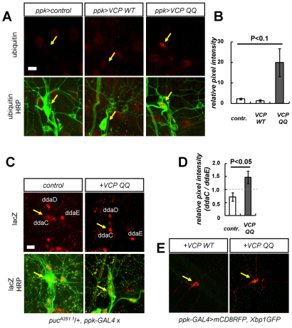
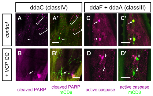
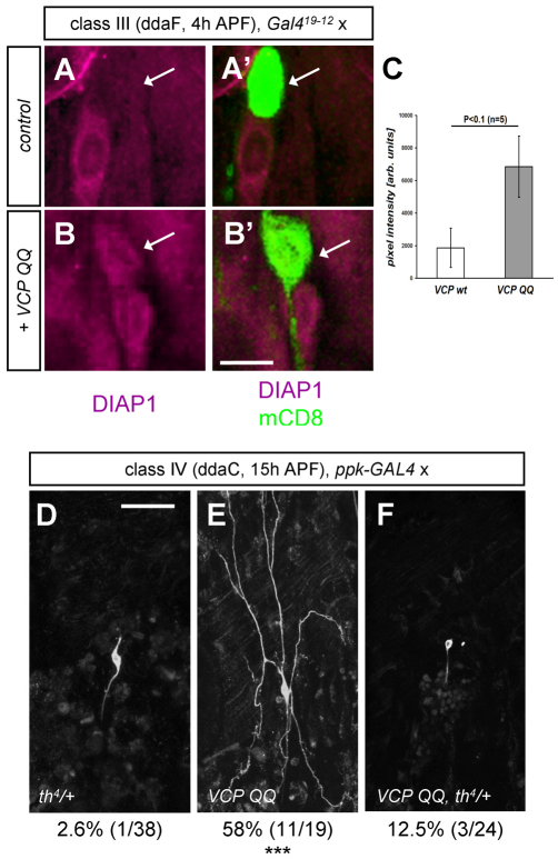
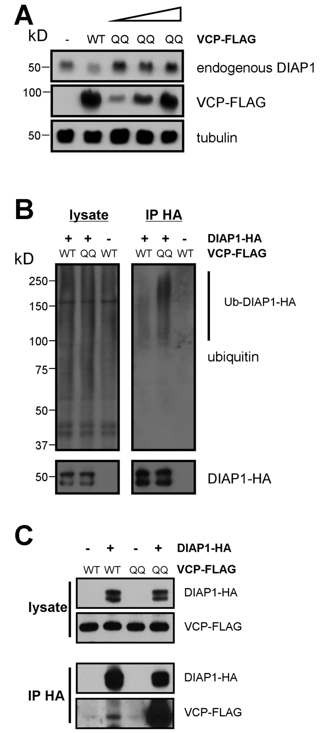
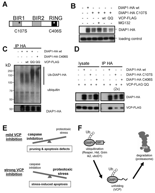
The regulated degeneration of axons or dendrites (pruning) and neuronal apoptosis are widely used during development to determine the specificity of neuronal connections. Pruning and apoptosis often share similar mechanisms; for example, developmental dendrite pruning of Drosophila class IV dendritic arborization (da) neurons is induced by local caspase activation triggered by ubiquitin-mediated degradation of the caspase inhibitor DIAP1. Here, we examined the function of Valosin-containing protein (VCP), a ubiquitin-selective AAA chaperone involved in endoplasmic reticulum-associated degradation, autophagy and neurodegenerative disease, in Drosophila da neurons. Strong VCP inhibition is cell lethal, but milder inhibition interferes with dendrite pruning and developmental apoptosis. These defects are associated with impaired caspase activation and high DIAP1 levels. In cultured cells, VCP binds to DIAP1 in a ubiquitin- and BIR domain-dependent manner and facilitates its degradation. Our results establish a new link between ubiquitin, dendrite pruning and the apoptosis machinery.


Figures

Fig. 1.
Fig. 1.
VCP inhibition affects dendrite morphology and cell viability of Drosophila larval da neurons. (A-D) VCP inhibition affects the morphology of class IV neurons. The indicated transgenes were expressed under the control of ppk-GAL4, and the class IV neuron ddaC was visualized by concomitant expression of membrane-bound GFP and imaged live at third instar. (A) Control (ppk-GAL4/+), (B) VCP RNAi (ppk-GAL4/UAS-VCP IR) and (C) VCP QQ (ppk-GAL4/UAS-VCP QQ). (D) Quantification of branching defects upon expression of VCP RNAi or VCP QQ. Data are shown as mean ± s.d., n=4; ***, P<0.0001 (Student's t-test). (E-H) No major effects of VCP inhibition on class III neuronal morphology. The indicated transgenes were expressed under Gal419-12, and the class III neurons ddaF and ddaA were imaged live at third instar. (E) Control (Gal419-12/+), (F) VCP RNAi (Gal419-12/UAS-VCP IR) and (G) VCP QQ (Gal419-12/UAS-VCP QQ). The insets show class III neuron dentrites at higher magnification for better comparison of spike density. (H) Quantification of spike numbers per 30 μm dendrite length for the indicated genotypes. Data are mean ± s.d., n=5. (I-K) Strong VCP inhibition causes severe morphological defects and lethality in class IV neurons. (I) ppk-GAL4/+, dvcp7-12/+ (heterozygous control). (J) Expression of VCP RNAi in a vcp heterozygous background (ppk-GAL4/UAS-VCP IR, dvcp7-12/+). (K) Two copies of UAS-VCP QQ (ppk-GAL4/2x UAS-VCP QQ). Arrows (J,K) indicate fragmenting dendrites. The number of dying or severely affected neurons for each genotype is indicated below each panel (***, P<0.0001; Fisher's exact test). Scale bars: 50 μm.
Fig. 2.
Fig. 2.
VCP inhibition induces proteotoxic stress in class IV neurons. (A,B) Ubiquitin staining of Drosophila third instar ppk neurons expressing different VCP transgenes. (A) The dorsal da neuron cluster of control animals and animals expressing wild-type VCP or VCP QQ in class IV neurons was stained with anti-ubiquitin antibodies (red). Genotypes from left to right: ppk-GAL4/+; ppk-GAL4/UAS-VCP wt; ppk-GAL4/UAS-VCP QQ. Dorsal da neurons were marked with anti-HRP antibodies (green); the class IV neuron ddaC is indicated by an arrow. (B) Quantification of the stainings in A. The relative pixel intensity of the ubiquitin signal of ddaC was compared with that of a neighboring class I da neuron (ddaE). Mean ± s.e.m., n=3. (C,D) VCP inhibition induces JNK activation in ppk neurons. (C) JNK activity in control ppk neurons or ppk neurons expressing VCP QQ was measured using a puc-lacZ (pucA251.1) reporter. JNK reporter activity was visualized by anti-β-galactosidase staining, and dorsal cluster da neurons were stained with anti-HRP antibodies. Genotypes: left, pucA251.1/+, ppk-GAL4/+; right, pucA251.1/+, ppk-GAL4/UAS-VCP QQ. The position of ddaC is indicated by an arrow. (D) Quantification of β-galactosidase staining shown in C. The quantification was performed as in B. Data are shown as mean ± s.d., n=3. (E) VCP inhibition induces endoplasmic reticulum (ER) stress. The UPR marker Xbp1-GFP was expressed in class IV neurons (labeled by mCD8::RFP). Wild-type VCP (left) does not induce visible GFP fluorescence (ppk-GAL4/UAS-mCD8::RFP, UAS-VCP wt, UAS-Xbp1-GFP), whereas expression of VCP QQ (right) induces a nuclear GFP signal indicative of ER stress in 70% of neurons (ppk-GAL4/UAS-mCD8::RFP, UAS-VCP QQ, UAS-Xbp1-GFP) (n=10 each). ddaC is indicated by an arrow.
Fig. 3.
Fig. 3.
VCP is required for dendrite pruning and apoptosis during Drosophila metamorphosis. Class IV neurons (ddaC) or class III neurons (ddaA and ddaF) expressing the indicated combinations of transgenes were imaged at 18 hours APF. (A-D) Effects of VCP inhibition on class IV neuron dendrite pruning. (A) Control (ppk-GAL4/+), (B) VCP RNAi (ppk-GAL4/UAS-VCP IR), (C) overexpression of wild-type VCP (ppk-GAL4/UAS-VCP wt) and (D) VCP QQ (ppk-GAL4/UAS-VCP QQ). The penetrance of pruning defects for each genotype is given under each panel (***, P<0.0001; Fisher's exact test). (E,F) Inducible VCP inhibition using ppk-GeneSwitch. Class IV neuron morphology was visualized with a ppk-CD4::tdTomato transgene (genotype ppk-GS/UAS-VCP QQ, ppk-CD4::tdTomato). VCP QQ expression was induced 24 hours before pupariation with (F) or without (E, control) 100 μM RU486. (G-K) VCP inhibition inhibits apoptosis of class III neurons during metamorphosis. (G) Control (Gal419-12/+), (H) class III neurons expressing p35 (Gal419-12/UAS-p35), (I) VCP RNAi (Gal419-12/UAS-VCP IR), (J) wild-type VCP (Gal419-12/UAS-VCP wt) and (K) VCP QQ (Gal419-12/UAS-VCP QQ). Control class III neurons or class III neurons expressing wild-type VCP have undergone apoptosis (G,J). Cell bodies of surviving class III neurons are marked by arrows (H,I,K). The number of surviving class III neurons is given below each panel (***, P<0.0001; Fisher's exact test). Scale bars: 50 μm.
Fig. 4.
Fig. 4.
VCP inhibition affects caspase activity during early metamorphosis. (A-B′) Caspase activity in class IV neurons was detected 4-6 hours APF with the reporter construct CD8::PARP::Venus. (A,A′) Control ddaC neuron (ppk-GAL4/UAS-CD8::Parp::Venus) and (B,B′) ddaC neuron expressing VCP QQ (ppk-GAL4/UAS-CD8::PARP::Venus, UAS-VCP QQ) (n=10 for each sample). The brackets (A,A′) indicate cleaved PARP in dendrites. (C-D′) Caspase activity in the class III neurons ddaA and ddaF (which is more dorsal) at 4 hours APF was detected with an antibody against activated caspases. (C,C′) Class III neurons overexpressing wild-type VCP (Gal419-12/UAS-VCP wt) and (D,D′) class III neurons expressing VCP QQ (Gal419-12/UAS-VCP QQ) (n=11 for each sample). Arrows (A-D′) indicate the positions of cell bodies (with weak caspase activity in or around the nucleus in B,B′). Scale bars: 50 μm in A-B′; 20 μm in C-D′.
Fig. 5.
Fig. 5.
Evidence that VCP affects DIAP1 during developmental apoptosis and pruning. (A-C) DIAP1 levels are upregulated in class III neurons during early metamorphosis (4 hours APF). (A,A′) DIAP1 levels in the class III neuron ddaF expressing wild-type VCP (Gal419-12/UAS-VCP wt). DIAP1 levels in the ddaF soma are lower than those in an adjacent cell (probably ddaC) and the cell body has rounded up. (B,B′) ddaF neuron expressing VCP QQ (Gal419-12/UAS-VCP QQ). DIAP1 levels are comparable to those in the adjacent cell and the ddaF soma retains its normal shape. (C) Quantification of normalized DIAP1 staining intensities in class III neurons expressing wild-type VCP or VCP QQ. Data are shown as mean ± s.d., n=5. (D-F) The DIAP1 mutant allele th4 is a suppressor of VCP during dendrite pruning. Class IV da neurons were imaged at 15 hours APF. (D) Class IV neuron in a th4/+ heterozygous background (th4/+, ppk-GAL4/+), (E) a class IV neuron expressing VCP QQ in the wild-type background (+/+, ppk-GAL4/UAS-VCP QQ) and (F) a class IV neuron expressing VCP QQ in a heterozygous DIAP1 mutant background (th4/+, ppk-GAL4/UAS-VCP QQ). The numbers of unpruned neurons are given below each panel. Scale bars: 10 μm in A-B′; 50 μm in D.
Fig. 6.
Fig. 6.
VCP is required for DIAP1 degradation in S2 cells. (A) VCP QQ expression leads to an accumulation of endogenous DIAP1. Drosophila S2 cells were transfected with wild-type VCP (0.8 μg) or increasing amounts of VCP QQ (0.2-0.8 μg), and cell extracts were probed with anti-DIAP1 antibodies. Tubulin served as a loading control. (B) Accumulation of ubiquitylated DIAP1 upon VCP inhibition. DIAP1-HA immunoprecipitates from cells expressing the indicated VCP constructs were blotted against ubiquitin. (C) Interactions between DIAP1 and VCP. DIAP1-HA and FLAG-tagged VCP constructs were expressed in S2 cells. HA immunoprecipitates (IP HA) were blotted with the indicated antibodies.
Fig. 7.
Fig. 7.
DIAP1 domains required for interaction with VCP. (A) Domain structure of DIAP1. The approximate positions of the mutations in C107S and C406S are indicated by asterisks. (B) Disrupton of the BIR1 domain destabilizes DIAP1. HA-tagged DIAP1 or DIAP1 C107S was expressed in Drosophila S2 cells and equal amounts of cell lysate were analyzed by SDS-PAGE. Where indicated, the proteasome inhibitor MG132 was added, or wild-type VCP or VCP QQ were co-expressed with DIAP1. The loading control was a cross-reactive band from a GAPDH antibody. (C) Effect of a RING finger mutation on the DIAP1 ubiquitylation status. HA-tagged DIAP1 or DIAP1 C406S was expressed in S2 cells together with VCP or VCP QQ, and DIAP1 was immunoprecipitated as in Fig. 6B. Immunoprecipitates were analyzed by blotting against HA and ubiquitin. (D) Effect of DIAP1 BIR1 and RING mutations on the interaction with VCP QQ. Wild-type DIAP1 or the BIR1 and RING mutants were co-transfected with VCP QQ and immunoprecipitated as in Fig. 6C. ‘2×’ indicates that twice as much BIR1 mutant immunoprecipitate was loaded to correct for the different expression levels. See also Fig. S3 in the supplementary material. (E) Model for the biphasic effects of VCP inhibition in neuronal viability, apoptosis and pruning. (F) Hypothetical mechanism for VCP-dependent DIAP1 degradation. The BIR1 domain stabilizes DIAP1 and necessitates VCP involvement to facilitate BIR1 domain unfolding and degradation. Ub, ubiquitin chain.

Similar articles

Cited by

References

    1. Beskow A., Grimberg K. B., Bott L. C., Salomons F. A., Dantuma N. P., Young P. (2009). A conserved unfoldase activity for the p97 AAA-ATPase in proteasomal degradation. J. Mol. Biol. 394, 732-746 - PubMed
    1. Brand A. H., Perrimon N. (1993). Targeted gene expression as a means of altering cell fates and generating dominant phenotypes. Development 118, 401-415 - PubMed
    1. Dietzl G., Chen D., Schnorrer F., Su K. C., Barinova Y., Fellner M., Gasser B., Kinsey K., Oppel S., Scheiblauer S., et al. (2007). A genome-wide transgenic RNAi library for conditional gene inactivation in Drosophila. Nature 448, 151-156 - PubMed
    1. Ditzel M., Wilson R., Tenev T., Zachariou A., Paul A., Deas E., Meier P. (2003). Degradation of DIAP1 by the N-end rule pathway is essential for regulating apoptosis. Nat. Cell Biol. 5, 467-473 - PubMed
    1. Dutta D., Bloor J. W., Ruiz-Gomez M., VijayRaghavan K., Kiehart D. P. (2002). Real-time imaging of morphogenetic movements in Drosophila using Gal4-UAS-driven expression of GFP fused to the actin-binding domain of moesin. Genesis 34, 146-151 - PubMed

Publication types

MeSH terms