. 2011 May;300(5):E877-85.
doi: 10.1152/ajpendo.00626.2010. Epub 2011 Feb 22.

Regulation of HIF-1{alpha} activity in adipose tissue by obesity-associated factors: adipogenesis, insulin, and hypoxia

Affiliations

Regulation of HIF-1{alpha} activity in adipose tissue by obesity-associated factors: adipogenesis, insulin, and hypoxia

Qing He et al. Am J Physiol Endocrinol Metab. 2011 May.

Abstract

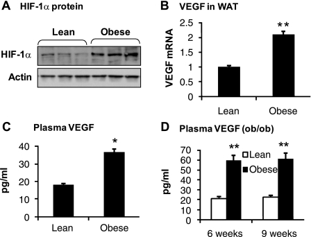
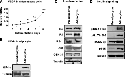
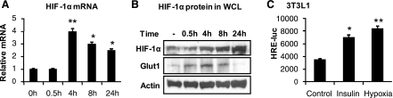
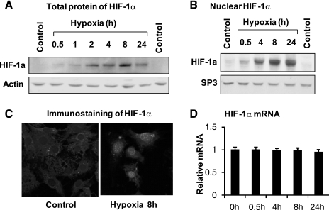
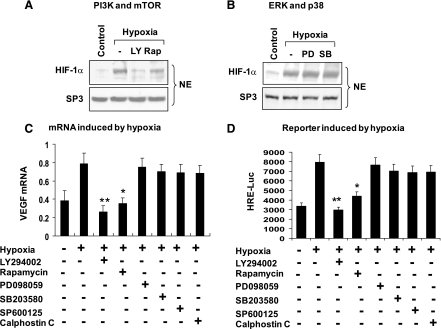
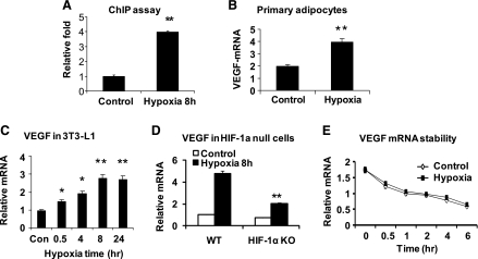
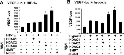
The transcription factor HIF-1α activity is increased in adipose tissue to contribute to chronic inflammation in obesity. However, its upstream and downstream events remain to be characterized in adipose tissue in obesity. We addressed this issue by investigating adipocyte HIF-1α activity in response to obesity-associated factors, such as adipogenesis, insulin, and hypoxia. In adipose tissue, both HIF-1α mRNA and protein were increased by obesity. The underlying mechanism was investigated in 3T3-L1 adipocytes. HIF-1α mRNA and protein were augmented by adipocyte differentiation. In differentiated adipocytes, insulin further enhanced HIF-1α in both levels. Hypoxia enhanced only HIF-1α protein, not mRNA. PI3K and mTOR activities are required for the HIF-1α expression. Function of HIF-1α protein was investigated in the regulation of VEGF gene transcription. ChIP assay shows that HIF-1α binds to the proximal hypoxia response element in the VEGF gene promoter, and its function is inhibited by a corepressor composed of HDAC3 and SMRT. These observations suggest that of the three obesity-associated factors, all of them are able to augment HIF-1α protein levels, but only two (adipogenesis and insulin) are able to enhance HIF-1α mRNA activity. Adipose tissue HIF-1α activity is influenced by multiple signals, including adipogenesis, insulin, and hypoxia in obesity. The transcriptional activity of HIF-1α is inhibited by HDAC3-SMRT corepressor in the VEGF gene promoter.


Figures

Fig. 1.
Fig. 1.
Hypoxia-inducible factor-1 (HIF-1) function indicated by plasma vascular endothelial growth factor (VEGF) in obese mice. A: HIF-1α protein in the adipose tissue. Tissue homogenizer was made from epididymal fat of dietary obese mice and examined for HIF-1α protein in a Western blot. B: VEGF mRNA in white adipose tissue (WAT) of dietary obese mice. mRNA was quantified with quantitative real-time PCR (qRT-PCR) in epididymal fat of mice on high-fat diet (HFD) for 12 wk. C: plasma VEGF protein in dietary obese mice. The protein was determined using an ELISA assay in samples collected from mice on HFD at 12 wk (n = 10). D: Plasma VEGF in ob/ob mice. The test was conducted at 6 and 9 wk of age (n = 7). In the bar graph, each data point represents means ± SE. *P < 0.05, **P < 0.001 (compared with control).
Fig. 2.
Fig. 2.
HIF-1α regulation by cell differentiation. A: VEGF mRNA during adipogenesis of 3T3-L1. Total mRNA was prepared from cells collected at times as indicated and quantified for VEGF mRNA by qRT-PCR. B: HIF-1α protein in the whole cell lysate of differentiated 3T3-L1 cells. The protein was determined in a Western blot. C: proteins in the insulin-signaling pathway. The proteins were determined in the whole cell lysate of differentiated 3T3-L1 cells in a Western blot. D: phosphorylation status of the signaling proteins. Phosphospecific antibodies were used in the assay of whole cell lysate, as described in materials and methods. In the chart, each data point represents means ± SE (n = 3). *P < 0.05, **P < 0.001 (compared with control). IRα and -β, insulin receptor-α and -β, respectively; IRS-1, insulin receptor substrate-1.
Fig. 3.
Fig. 3.
HIF-1α regulation by insulin in differentiated 3T3-L1 cells. A: HIF-1α mRNA in cells treated with insulin (100 nM). B: HIF-1α and glucose transporter 1 (Glut1) proteins in whole cell lysate of differentiated 3T3-L1 adipocytes treated with insulin. C: HIF-1 function in hypoxia response element-luciferase (HRE-luc) reporter. Luciferase assay was performed in 3T3-L1 adipocytes that were transiently transfected with the reporter and treated with insulin for 16 h. Hypoxia treatment is a positive control. In the bar graph, each data point represents means ± SE (n = 3). *P < 0.05, **P < 0.001 (compared with control).
Fig. 4.
Fig. 4.
Regulation of HIF-1α by hypoxia. A: HIF-1α total protein. The protein was determined in the whole cell lysate in Western blot. Differentiated 3T3-L1 cells were treated with hypoxia (1% oxygen) for different times as indicated. B: nuclear HIF-1α protein. The nuclear extract was made from cells treated with hypoxia for different times and quantified for HIF-1α protein in a Western blot. Specific protein 3 (SP3) is a loading control. C: immunofluorescent staining of HIF-1α protein in 3T3-L1 adipocytes. D: mRNA of HIF-1α. The mRNA was determined in differentiated 3T3-L1 cells after hypoxia treatment. D: each data point represents means ± SE (n = 3).
Fig. 5.
Fig. 5.
Signaling for HIF-1α elevation. A: phosphatidylinositol 3-kinase (PI3K) and mammalian target of rapamycin (mTOR) in the control of HIF-1α activity. Differentiated 3T3-L1 cells were serum starved and pretreated with LY-294002 (LY) and rapamycin (Rap) for 0.5 h and then treated with hypoxia for 8 h. The HIF-1α protein was examined in the nuclear extract in a Western blot. B: ERK and p38. The nuclear HIF-1α was determined in differentiated 3T3-L1 cells treated with PD-098059 (PD) or SB-203580 (SB) for 0.5 h. C: inhibition of HIF-1 function by kinase inhibitors. Differentiated 3T3-L1 cells were pretreated with kinase inhibitors such as LY (PI3K), Rap (mTOR), PD (MEK/ERK), SB (p38), SP-600125 (JNK), and calphostin C (PKC) for 0.5 h, followed by hypoxia treatment. VEGF mRNA was examined after hypoxia treatment for 8 h. D: HRE-luc assay. The HRE-luc reporter was transfected into 293 cells and induced with hypoxia after the inhibitor treatment for 0.5 h. The luciferase activity was measured after hypoxia treatment for 8 h. In the bar graph, each data point represents means ± SE. *P < 0.05, **P < 0.001 (n = 3). NE, nuclear extract.
Fig. 6.
Fig. 6.
HIF-1α in transcriptional expression of VEGF. A: chromatin immunoprecipitation (ChIP) assay for HIF-1α in the VEGF gene promoter. The assay was conducted in differentiated 3T3-L1 adipocytes after hypoxia treatment for 8 h. B: hypoxia induction of VEGF mRNA in primary adipocytes. C: VEGF induction in 3T3-L1 adipocytes. D: VEGF mRNA in HIF-1α-knockout (KO) cells. mouse embryonic fibroblasts from HIF-1α-KO mice were treated with hypoxia for 8 h in the experiment. E: VEGF mRNA stability in differentiated 3T3-L1 adipocytes. In the bar graph, each data point represents means ± SE. *P < 0.05, **P < 0.001 (n = 3–5). WT, wild type.
Fig. 7.
Fig. 7.
Inhibition of HIF-1α function by histone deacetylase (HDAC)3 and silencing mediator for retinoic and thyroid hormone receptors (SMRT). Functions of HDAC1, HDAC2, HDAC3, SMRT, and nuclear corepressor (NCoR) in the regulation of HIF-1 activity were examined using VEGF-luc reporter in the 293 cells. The corepressor proteins were knocked down with RNAi that was expressed from plasmid vectors cotransfected. The reporter activity was induced by either HIF-1α overexpression or hypoxia treatment. After transfection for 24 h, the cells were treated with hypoxia for 8 h in serum-free medium. A: VEGF-luc activity induced by HIF-1α expression. B: VEGF-luc activity induced by hypoxia. In the bar graph, each data point represents means ± SE. *P < 0.05 (n = 3).

References

    1. Arany Z, Foo SY, Ma Y, Ruas JL, Bommi-Reddy A, Girnun G, Cooper M, Laznik D, Chinsomboon J, Rangwala SM, Baek KH, Rosenzweig A, Spiegelman BM. HIF-independent regulation of VEGF and angiogenesis by the transcriptional coactivator PGC-1alpha. Nature 451: 1008–1012, 2008 - PubMed
    1. Arany Z, Huang LE, Eckner R, Bhattacharya S, Jiang C, Goldberg MA, Bunn HF, Livingston DM. An essential role for p300/CBP in the cellular response to hypoxia. Proc Natl Acad Sci USA 93: 12969–12973, 1996 - PMC - PubMed
    1. Bedogni B, Welford SM, Cassarino DS, Nickoloff BJ, Giaccia AJ, Powell MB. The hypoxic microenvironment of the skin contributes to Akt-mediated melanocyte transformation. Cancer Cell 8: 443–454, 2005 - PubMed
    1. Bradbury D, Clarke D, Seedhouse C, Corbett L, Stocks J, Knox A. Vascular endothelial growth factor induction by prostaglandin E2 in human airway smooth muscle cells is mediated by E prostanoid EP2/EP4 receptors and SP-1 transcription factor binding sites. J Biol Chem 280: 29993–30000, 2005 - PubMed
    1. Bråkenhielm E, Cao R, Gao B, Angelin B, Cannon B, Parini P, Cao Y. Angiogenesis inhibitor, TNP-470, prevents diet-induced and genetic obesity in mice. Circ Res 94: 1579–1588, 2004 - PubMed

Publication types

MeSH terms