. 2011 May 12;117(19):5057-66.
doi: 10.1182/blood-2010-08-300145. Epub 2011 Feb 22.

Prdm16 is a physiologic regulator of hematopoietic stem cells

Affiliations

Prdm16 is a physiologic regulator of hematopoietic stem cells

Francesca Aguilo et al. Blood. .

Abstract

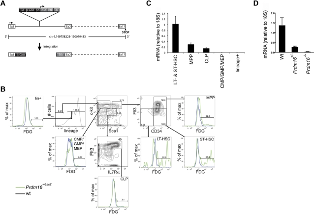
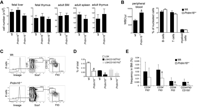
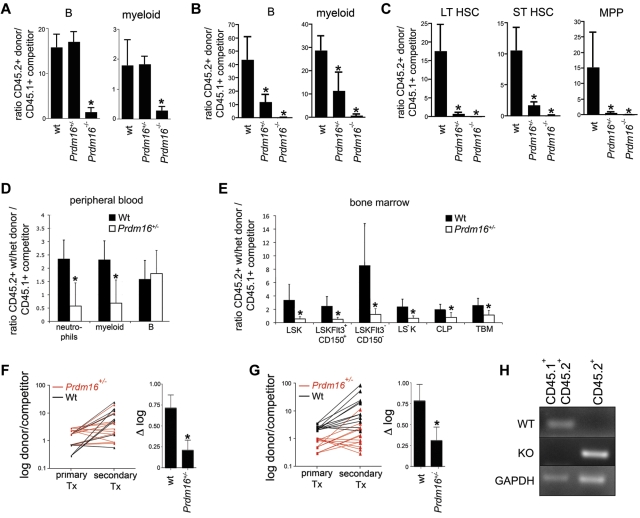
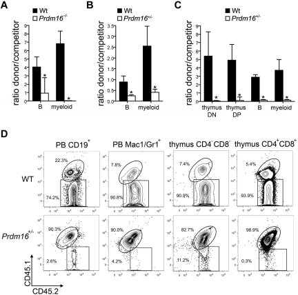
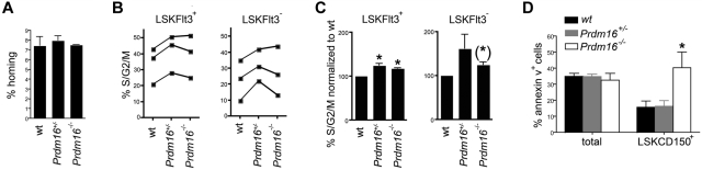
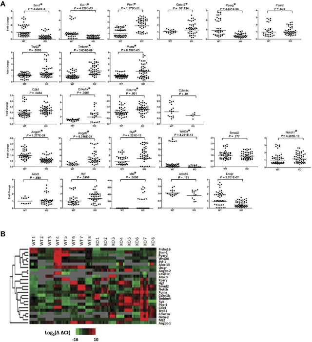
Fetal liver and adult bone marrow hematopoietic stem cells (HSCs) renew or differentiate into committed progenitors to generate all blood cells. PRDM16 is involved in human leukemic translocations and is expressed highly in some karyotypically normal acute myeloblastic leukemias. As many genes involved in leukemogenic fusions play a role in normal hematopoiesis, we analyzed the role of Prdm16 in the biology of HSCs using Prdm16-deficient mice. We show here that, within the hematopoietic system, Prdm16 is expressed very selectively in the earliest stem and progenitor compartments, and, consistent with this expression pattern, is critical for the establishment and maintenance of the HSC pool during development and after transplantation. Prdm16 deletion enhances apoptosis and cycling of HSCs. Expression analysis revealed that Prdm16 regulates a remarkable number of genes that, based on knockout models, both enhance and suppress HSC function, and affect quiescence, cell cycling, renewal, differentiation, and apoptosis to various extents. These data suggest that Prdm16 may be a critical node in a network that contains negative and positive feedback loops and integrates HSC renewal, quiescence, apoptosis, and differentiation.


Figures

Figure 1
Figure 1
Expression of Prdm16 in adult BM. (A) Schematic representation of the genomic Prdm16 region in Prdm16Gt(OST67423)Lex (Prdm16−/−) mice. Adapted from Zambrowicz et al with permission. (B) Expression of LacZ, measured by flow cytometry after staining with fluorescein di-D-β-galactopyranoside, in the BM of Prdm16Gt(OST67423)Lex/+ mice. CMP indicates common myeloid progenitor; GMP, granulocyte/macrophage progenitor; MEP, megakaryocyte erythroid progenitor; LT-HSC, long-term hematopoietic stem cell; and ST-HSC, short-term hematopoietic stem cell. (C) Expression of Prdm16 mRNA in HSCs (LSKFlt3, composed of ST- and LT-HSCs), MPPs, CLPs, and lineage+ cells (n = 3). (D) Expression of Prdm16 mRNA in brain from Prdm16−/−, Prdm16+/−, and wt embryos.
Figure 2
Figure 2
Hematologic profile of Prdm16-deficient mice. (A) Cellularity of FL and thymus, and of adult BM, thymus, and spleen in Prdm16-deficient mice (n = 3–5). (B) WBC count (left panel) and lineage distribution (measured by flow cytometric analysis of CD19, Thy1, and Mac1/Gr1) in the PB of adult Prdm16+/− and wt mice (n = 5). *P = .0006. (C) Representative example of flow cytometric analysis of the fraction of LSK HSPCs, LSK CD150Flt3+ MPPs, and LSK CD150+Flt3 HSCs in E15 Prdm16−/− and wt FL cells. (D) Frequency of the populations in panel C in E13.5 to E15.5 FL from Prdm16−/− and wt embryos. Frequencies obtained as the percentage of cells in doublet discriminated scatter (n = 4-6). *P < .05, one-way analysis of variance. (E) Frequencies of subpopulations of LSK cells in the BM of Prdm16+/− mice or wt littermates. Lineage cocktail included anti–CD41 and CD48 (n = 8). *P < .03.
Figure 3
Figure 3
Function of Prdm16–deficient FL and BM cells. (A) Ratio between donor (Prdm16−/−, Prdm16+/−, or wt FL cells) and competitor (T cell–depleted CD45.1+ or CD45.1+CD45.2+ B6 BM cells) cells among PB myeloid and B cells 12 weeks after transplantation of 0.5 × 10⁶ cells of each population into lethally irradiated B6.129F1 (CD45.2+) mice (n = 3 for wt, n = 12 for Prdm16+/−, n = 14 for Prdm16−/−). *P < .01, one-way analysis of variance. (B-C) Ratio between donor (Prdm16−/−, Prdm16+/−, or wt FL cells) and competitor (T cell–depleted CD45.1+ B6 BM cells) cells among PB myeloid and B cells (B) and among BM LT-HSCs, ST-HSCs, and MPPs (C) 20 weeks after transplantation as in panel A. (D-E) Competitive repopulation of 2 × 10⁶ wt or Prdm16+/− BM cells (CD45.2+) with 2 × 10⁶ C57BL/6 (CD45.1+) BM cells in B6.129F1 recipients after 12 to 16 weeks. Data presented as donor/competitor ratio within a specific compartment in PB (D) and BM (E) (n = 6). *P < .02. TBM indicates total BM. (F-G) Shift in donor/competitor ratio within B cells (F) and myeloid cells (G) after serial transplantation of 2 × 10⁶ BM cells from primary recipients into lethally irradiated secondary B6.129F1 CD45.2+ recipients 12 to 14 weeks after primary transplantation. The right hand panels of panels F and G show the difference in log(donor/competitor) between primary and secondary recipients (n = 12 secondary recipients for each genotype). *P = .07 for myeloid cells. *P = .01 for B cells. Tx indicates transplantation. (H) Genomic PCR for the wt and mutant alleles in the myeloid and B cells of the spleens of recipients of CD45.2+ Prdm16−/− cells and CD45.1+CD45.2+ B6 competitor cells.
Figure 4
Figure 4
Function of Prdm16-deficient HSPCs. (A-C) Ratio between donor (300 Prdm16−/− or wt FL LSK cells (n = 4) (A), 500 Prdm16+/− or wt adult BM LSK cells (n = 9) (B), or 300 Prdm6+/− or wt adult BM LSKCD34Flt3 cells (n = 4) (C) and competitor (0.5 × 10⁶ T cell-depleted CD45.1+ or CD45.1+CD45.2+ B6 BM cells) cells among PB myeloid and B cells, and (C) thymic CD4+CD8+ double-positive and CD4CD8 double-negative developing T cells, 10 to 15 weeks after transplantation into lethally irradiated B6.129F1 (CD45.2+) mice. *P < .01. (D) Representative example of donor (CD45.2+) and competitor (CD45.1+CD45.2+) reconstitution in doublet discriminated PB CD19+ B and Mac1+Gr1+ myeloid cells, and in thymic CD4CD8 double-negative and CD4+CD8+ double-positive cells after competitive transplantation with wt or Prdm16+/− adult BM LT-HSCs.
Figure 5
Figure 5
Effects of Prdm16 on homing, apoptosis, and cell cycling. (A) Homing of Prdm16−/−, Prdm16+/−, and wt E16 FL cells after transplantation into lethally irradiated CD45.1+ recipients (n = 3). (B) Percentage of FL LSKFlt3+ and LSKFlt3 cells from Prdm16−/−, Prdm16+/−, and wt mice in S/G2/M phase of the cell cycle as measured by 4,6-diamidino-2-phenylindole staining. Each experiment contained 2 to 4 embryos per genotype. (C) Fraction of FL LSKFlt3+ and LSKFlt3 cells from Prdm16−/−, Prdm16+/−, and wt mice in S/G2/M phase normalized to the values of wt littermate embryos (n = 3 litters). *P < .05. (*)P = .08. (D) Percentage of apoptotic total FL and LSKCD150+ FL cells in Prdm16−/−, Prdm16+/−, and wt embryos (n = 3 for each genotype). *P = .05, one-way ANOVA.
Figure 6
Figure 6
Expression analysis of Prdm16−/− and wt FL HSCs. (A) Scatter dot plots of the expression of the genes, as measured using Fluidigm multiplex quantitative PCR, indicated on top of each panel in 8 groups of 5 Prdm16−/− or wt FL LSKCD150+ HSCs. *P < .002, threshold required after Bonferroni correction. All experimental replicates of the amplification step and the microfluidic quantitative PCR chip are shown. Undetectable expression is defined by a Ct value of 35 or greater. (B) Heat map of the expression levels for Prdm16 and 24 genes shown in panel A.

Comment in

Similar articles

Cited by

References

    1. Orkin SH, Zon LI. Hematopoiesis: an evolving paradigm for stem cell biology. Cell. 2008;132(4):631–644. - PMC - PubMed
    1. Ernst P, Fisher JK, Avery W, et al. Definitive hematopoiesis requires the mixed-lineage leukemia gene. Dev Cell. 2004;6(3):437–443. - PubMed
    1. McMahon KA, Hiew SY, Hadjur S, et al. Mll has a critical role in fetal and adult hematopoietic stem cell self-renewal. Cell Stem Cell. 2007;1(3):338–345. - PubMed
    1. Jude CD, Climer L, Xu D, Artinger E, Fisher JK, Ernst P. Unique and independent roles for MLL in adult hematopoietic stem cells and progenitors. Cell Stem Cell. 2007;1(3):324–337. - PMC - PubMed
    1. Goyama S, Yamamoto G, Shimabe M, et al. Evi-1 is a critical regulator for hematopoietic stem cells and transformed leukemic cells. Cell Stem Cell. 2008;3(2):207–220. - PubMed

Publication types

MeSH terms