Review
. 2011 Mar-Apr;5(2):199-206.
doi: 10.4161/cam.5.2.15081. Epub 2011 Mar 1.

Emerging role for ERM proteins in cell adhesion and migration

Affiliations
Review

Emerging role for ERM proteins in cell adhesion and migration

Monique Arpin et al. Cell Adh Migr. 2011 Mar-Apr.

Abstract

The highly related ERM (Ezrin, Radixin, Moesin) proteins provide a regulated linkage between the membrane and the underlying actin cytoskeleton. They also provide a platform for the transmission of signals in responses to extracellular cues. Studies in different model organisms and in cultured cells have highlighted the importance of ERM proteins in the generation and maintenance of specific domains of the plasma membrane. A central question is how do ERM proteins coordinate actin filament organization and membrane protein transport/stability with signal transduction pathways to build up complex structures? Through their interaction with numerous partners including membrane proteins, actin cytoskeleton and signaling molecules, ERM proteins have the ability to organize multiprotein complexes in specific cellular compartments. Likewise, ERM proteins participate in diverse functions including cell morphogenesis, endocytosis/exocytosis, adhesion and migration. This review focuses on aspects still poorly understood related to the function of ERM proteins in epithelial cell adhesion and migration.


Figures
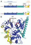
Figure 1
Figure 1
(A) Domain organization of human ezrin and Sfmoesin. All ERM proteins display similar domain organization. They are composed of a globular N-terminal domain (FERM domain) followed by a domain rich in α-helices, a linker region and the C-terminal domain. The F-actin binding site has been mapped to the last 30 carboxy-terminal aminoacids. Tyrosines 145 and 477 are phosphorylated by Src kinases downstream growth factor stimulation., These two tyrosines are not conserved in Sfmoesin. (B) Structure of full-length Spodoptera frugiperda moesin; protein data bank (PDB) identifier 2I1J. The three lobes that composed the FERM domain (F1, F2 and F3) are colored in dark blue, the α-helical domain in turquoise, the linker region in red and the C-terminal domain in yellow. In the closed state, the α-helical region and the C-terminal domain make extensive interactions with the FERM domain that mask the ligand binding sites. IP3 binds a basic cleft between domains F1 and F3 of the free FERM domain. Phosphorylation of threonine 556, highlighted in green, weakens the interaction between the FERM and the C-terminal domains.
Figure 2
Figure 2
Ezrin is an essential component in HGF-induced cell scattering. Upper part: I: HGF stimulation induces morphological changes including breakdown of the microvilli (II), cell flattening (II), cell-cell contact dissociation and increased cell migration (III). Lower part: At the molecular level, stimulation of the Met receptor activates multiple intracellular signaling pathways. (I) Following HGF stimulation, ezrin is recruited to the lateral membrane where it can interact with the adhesion molecule CD44 with concomitant phosphorylation on tyrosine residues. Phosphorylated tyrosine can interact with SH2-domain containing proteins such as the Fes kinase. Fes is recruited and activated to cell-cell contacts by ezrin phosphorylated at tyrosine 477. (II) Phosphorylation of regulatory cell junction proteins by activated Fes can trigger the dissociation of cell-cell contacts. (III) Activated Rac downstream of ezrin is involved in the epithelial to mesenchymal transition by regulating actin cytoskeleton remodeling and E-cadherin trafficking.

Similar articles

Cited by

References

    1. Zaidel-Bar R, Geiger B. The switchable integrin adhesome. J Cell Sci. 2010;123:1385–1388. - PMC - PubMed
    1. Sato N, Funayama N, Nagafuchi A, Yonemura S, Tsukita S, Tsukita S. A gene family consisting of ezrin, radixin and moesin. Its specific localization at actin filament/plasma membrane association sites. J Cell Sci. 1992;103:131–143. - PubMed
    1. Turunen O, Wahlström T, Vaheri A. Ezrin has a COOH-terminal actin-binding site that is conserved in the ezrin protein family. J Cell Biol. 1994;126:1445–1453. - PMC - PubMed
    1. Algrain M, Turunen O, Vaheri A, Louvard D, Arpin M. Ezrin contains cytoskeleton and membrane binding domains accounting for its proposed role as a membrane-cytoskeletal linker. J Cell Biol. 1993;120:129–139. - PMC - PubMed
    1. Gary R, Bretscher A. Ezrin self-association involves binding of an N-terminal domain to a normally masked C-terminal domain that includes the F-actin binding site. Mol Biol Cell. 1995;6:1061–1075. - PMC - PubMed

Publication types

MeSH terms

LinkOut - more resources