Review
. 2011 Apr;7(4):229-36.
doi: 10.1038/nrneurol.2011.21. Epub 2011 Feb 22.

White matter lesions in Parkinson disease

Affiliations
Review

White matter lesions in Parkinson disease

Nicolaas I Bohnen et al. Nat Rev Neurol. 2011 Apr.

Abstract

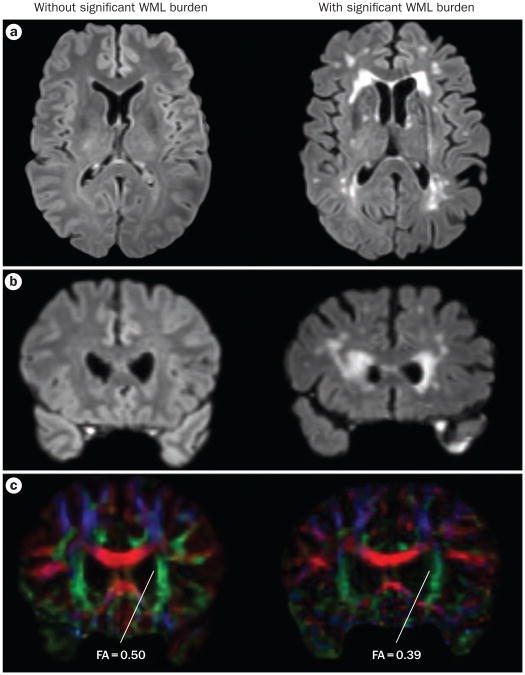
Pure vascular parkinsonism without evidence of nigral Lewy body pathology may occur as a distinct clinicopathological entity, but a much more frequent occurrence is the comorbid presence of age-associated white matter lesions (WMLs) in idiopathic Parkinson disease (PD). WMLs are associated with motor and cognitive symptoms in otherwise normal elderly individuals. Comorbid WMLs are, therefore, expected to contribute to clinical symptoms in PD. Studies of WMLs in PD differ with regard to methods of assessment of WML burden and the patient populations selected for analysis, but converging evidence suggests that postural stability and gait motor functions are predominantly affected. WMLs are described to contribute to dementia in Alzheimer disease, and emerging but inconclusive evidence indicates similar effects in PD. In this article, we review the literature addressing the occurrence and impact of WMLs in PD, and suggest that WMLs may exacerbate or contribute to some motor and cognitive deficits associated with PD. We review existing and emerging methods for studying white matter pathology in vivo, and propose future research directions.


Conflict of interest statement

Competing interests: The authors declare no competing interests.

Figures

Figure 1
Figure 1
Venn diagram showing overlap between idiopathic PD and WMLs of normal aging. Normal aging is accompanied by nigrostriatal degeneration, and the presence of WMLs has been associated with mild parkinsonian symptoms in elderly individuals without PD. Abbreviations: PD, Parkinson disease; WMLs, white matter lesions.
Figure 2
Figure 2
Imaging WMLs. Comparison of DTI and FLAIR MRI of PD patients without (left panels) and with (right panels) significant WML burden. a | Transaxial slices of the FLAIR image sets. Hyperintensities, especially in the frontal ventricular horns, are prominent in the patient on the right, and absent in the patient on the left. b | Coronal slices of the FLAIR image sets, taken near the anterior border of the lateral ventricles. c | Coronal DTI slices, taken at the same level as the coronal FLAIR images. Degradation of the anterior aspect of the corona radiata is apparent in the patient with significant FLAIR hyperintensities. FA is also reduced in this area. FA values are also slightly reduced in the patient without significant FLAIR hyperintensities, indicating decreased white matter microstructural integrity. These findings are consistent with altered diffusion in the frontal lobes, as observed in DTI studies in patients with PD., Abbreviations: DTI, diffusion tensor imaging; FA, fractional anisotropy; FLAIR, fluid-attenuated inversion recovery; WML, white matter lesion.
Figure 3
Figure 3
Monoaminergic and cholinergic pathways that can be disrupted by white matter lesions. Sagittal brain MRI slice showing the deep forebrain where subcortical monoaminergic and cholinergic axons pass close to the ventricles before fanning out to the cortex. The dopaminergic mesofrontal and limbic cortical pathways (green) originate from the ventral tegmental area. Noradrenergic cortical pathways (red) originate from the locus coeruleus. Serotonergic cortical pathways originate from the raphe nuclei (blue). Cholinergic projections originating from the nucleus basalis of Meynert are shown in black. Periventricular frontal white matter lesions are strategically located to disrupt these ascending neuromodulator system projections.

References

    1. Murray ME, et al. Functional impact of white matter hyperintensities in cognitively normal elderly subjects. Arch Neurol. 2010;67:1379–1385. - PMC - PubMed
    1. Baloh RW, Yue Q, Socotch TM, Jacobson KM. White matter lesions and disequilibrium in older people. I. Case–control comparison. Arch Neurol. 1995;52:970–974. - PubMed
    1. Gunning-Dixon FM, Raz N. The cognitive correlates of white matter abnormalities in normal aging: a quantitative review. Neuropsychology. 2000;14:224–232. - PubMed
    1. Baezner H, et al. Association of gait and balance disorders with age-related white matter changes: the LADIS study. Neurology. 2008;70:935–942. - PubMed
    1. Novak V, et al. White matter hyperintensities and dynamics of postural control. Magn Reson Imaging. 2009;27:752–759. - PMC - PubMed

Publication types

MeSH terms