. 2011 Dec;35(12):1530-8.
doi: 10.1038/ijo.2011.12. Epub 2011 Feb 22.

Suppression of inflammation-associated factors by indole-3-carbinol in mice fed high-fat diets and in isolated, co-cultured macrophages and adipocytes

Affiliations
Free PMC article

Suppression of inflammation-associated factors by indole-3-carbinol in mice fed high-fat diets and in isolated, co-cultured macrophages and adipocytes

H-P Chang et al. Int J Obes (Lond). 2011 Dec.
Free PMC article

Erratum in

  • Int J Obes (Lond). 2013 Feb;37(2):324

Abstract

Aims: This study investigated the effects of indole-3-carbinol (I3C), a compound from cruciferous vegetables, on various parameters related to obesity, in particular, the parameters of infiltration by macrophages and of inflammatory cytokines expressed during the co-culture of adipocytes and macrophages.

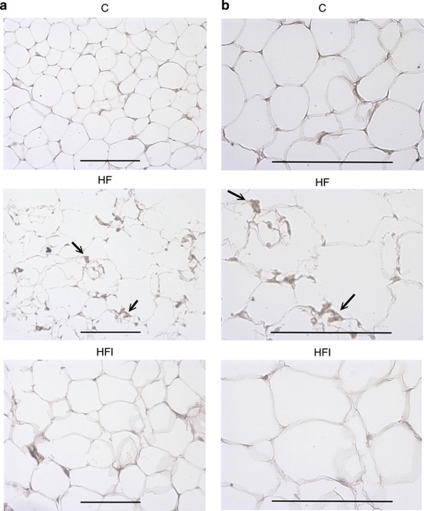
Methods: Male C57BL/6 mice were fed with a control diet (C group), high-fat diet (HF group) and HF+5 mg kg(-1) I3C (HFI group). The I3C was intraperitoneally injected (HFI group) for 12 weeks. Epididymal adipose tissue (AT) was collected and stained for F4/80, a marker of macrophages.

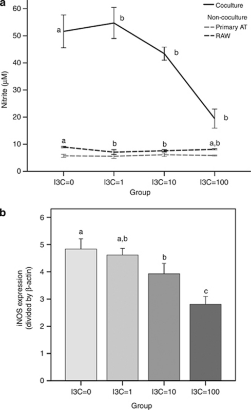
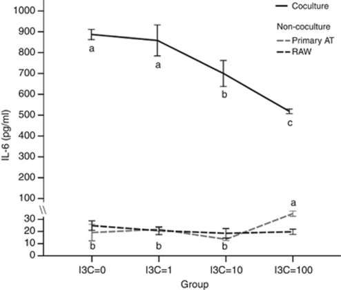
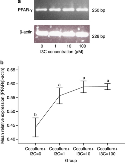
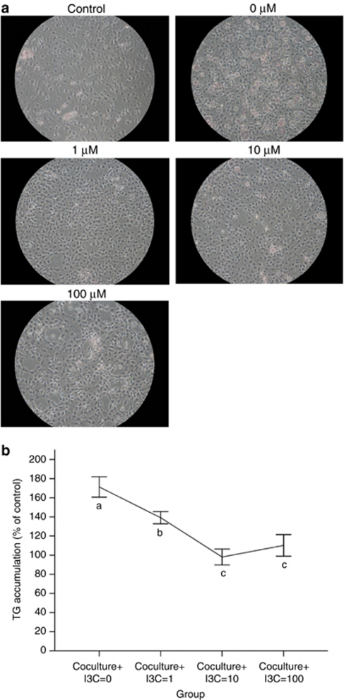
Results: The immunohistochemical staining for F4/80 indicated a greater presence of macrophages in the HF group than in AT from the control and HFI groups. Furthermore, I3C treatment, in an in vitro cell culture system, decreased expression of inducible nitric oxide synthase (iNOS), decreased nitrite content and enhanced expression of peroxisome proliferator-activated receptor (PPAR-γ). Moreover, in vitro cell culture studies revealed that I3C inhibited intracellular lipid accumulation in hypertrophied adipocytes. In macrophage and primary adipocyte co-culture, I3C inhibited expression of interleukin-6 (IL-6).

Conclusions: In vivo treatment with I3C reduced the infiltration of macrophages in AT, and in vitro addition of I3C to co-cultured macrophages and adipocytes reduced nitrite production and IL-6 expression. With cultures of adipocytes only, I3C inhibited accumulation of intracellular lipid, either by disrupting differentiation, or by directly inhibiting triglyceride synthesis.


Figures

Figure 1
Figure 1
Effects of I3C on F4/80 expression in epididymal adipose tissue (AT), × 200 magnification (a) and × 400 magnification (b), in animals fed different diets for 12 weeks (C, control diet; HF, high-fat diet; HFI, high-fat diet+intraperitoneally administered I3C). Immunohistochemistry detected a macrophage-specific antigen (F4/80) in epididymal AT from high-fat diet-induced obese mice. The black arrows showed abundant macrophages within AT. Macrophages were observed both near blood vessels and among large adipocytes. The scale bar represents 100 μm.
Figure 2
Figure 2
Effects of I3C on nitrite content and inducible nitric oxide synthase (iNOS) mRNA expression in co-culture of primary adipocytes and macrophages. Differentiated adipocytes were co-cultured with RAW 264.7 macrophages (1 × 10⁶ cells per well) or the cells were cultured alone (a) and cells were treated with I3C (μ) for 24 h. Nitrite levels were spectrophotometrically measured with Griess reagent. Expression of iNOS mRNA (b) was measured by reverse transcriptase polymerase chain reaction. Values are the mean±s.d. from six measurements. Values with different superscript letters (a, b and c) indicate significant differences between groups (P<0.05).
Figure 3
Figure 3
Effects of I3C on interleukin-6 (IL-6) in co-culture of primary adipocytes and macrophages. Differentiated adipocytes were co-cultured with RAW 264.7 macrophages (1 × 10⁶ cells per well), and cells were treated with I3C (μ) for 24 h. IL-6 contents in the co-culture model, primary adipocytes alone and RAW 264.7 macrophages alone, were measured by ELISA. Values are the mean±s.d. from six measurements. Values with different superscript letters (a, b and c) indicate significant differences between groups (P<0.05).
Figure 4
Figure 4
Effects of I3C on peroxisome proliferator-activated receptor (PPAR-γ) mRNA in co-culture of primary adipocytes and macrophages. Differentiated adipocytes were co-cultured with RAW 264.7 macrophages (1 × 10⁶ cells per well), and cell treated with I3C for 24 h. Differentiated adipocytes were treated with ethanol (0 μ I3C), 1 μ I3C, 10 μ I3C or 100 μ I3C, as indicated. The expression of PPAR-γ mRNAs was determined by reverse transcriptase polymerase chain reaction. The total cellular RNA was isolated and reverse-transcribed to cDNA (a). The relative expression of PPAR-γ under different concentrations of I3C was summarized (b). These experiments were repeated three times. Values with different superscript letters (a and b) indicate significant differences between groups (P<0.05).
Figure 5
Figure 5
Effects of I3C on intracellular lipid accumulation (a) and triglyceride accumulation (TG) content (b) in 3T3-L1 adipocytes. 3T3-L1 were differentiated by culturing with adipogenic agent. Cells were treated with various concentrations of I3C for 72 h. Cells were stained with oil red O. TG contents were quantified by a commercial assay kit, as described in Materials and Methods. These experiments were repeated three times, and similar results were obtained. Control: undifferentiated 3T3-L1 preadipocytes; 0 (0 μ I3C): differentiated 3T3-L1 adipocytes treated with ethanol; 1–100 μ: differentiated adipocytes treated with 1–100 μ I3C. Values are the mean±s.d. from three measurements. Values with different superscript letters (a, b, and c) indicate significant differences between groups (P<0.05).

Similar articles

Cited by

References

    1. Ghanim H, Aljada A, Hofmeyer D, Syed T, Mohanty P, Dandona P. Circulating mononuclear cells in the obese are in a proinflammatory state. Circulation. 2004;110:1564–1571. - PubMed
    1. Xu H, Barnes GT, Yang Q, Tan G, Yang D, Chou CJ, et al. Chronic inflammation in fat plays a crucial role in the development of obesity-related insulin resistance. J Clin Invest. 2003;112:1821–1830. - PMC - PubMed
    1. Olshansky SJ, Passaro DJ, Hershow RC, Layden J, Carnes BA, Brody J, et al. Ludwig, a potential decline in life expectancy in the United States in the 21st century. N Engl J Med. 2005;352:1138–1145. - PubMed
    1. Johnson JL, Newby AC. Macrophage heterogeneity in atherosclerotic plaques. Curr Opin Lipidol. 2009;20:370–378. - PMC - PubMed
    1. Zeyda M, Stulnig TM. Obesity, inflammation, and insulin resistance--a mini-review. Gerontology. 2009;55:379–386. - PubMed

MeSH terms