. 2011 Aug;19(8):1457-67.
doi: 10.1038/mt.2011.10. Epub 2011 Feb 22.

A peptide-based vector for efficient gene transfer in vitro and in vivo

Affiliations

A peptide-based vector for efficient gene transfer in vitro and in vivo

Taavi Lehto et al. Mol Ther. 2011 Aug.

Abstract

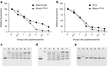
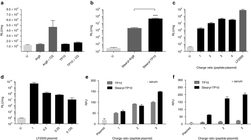
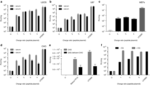
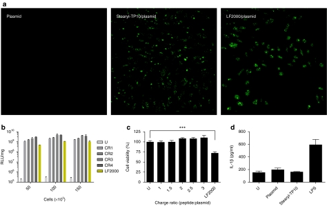
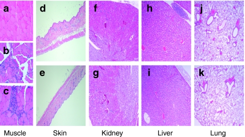
Finding suitable nonviral delivery vehicles for nucleic acid-based therapeutics is a landmark goal in gene therapy. Cell-penetrating peptides (CPPs) are one class of delivery vectors that has been exploited for this purpose. However, since CPPs use endocytosis to enter cells, a large fraction of peptides remain trapped in endosomes. We have previously reported that stearylation of amphipathic CPPs, such as transportan 10 (TP10), dramatically increases transfection of oligonucleotides in vitro partially by promoting endosomal escape. Therefore, we aimed to evaluate whether stearyl-TP10 could be used for the delivery of plasmids as well. Our results demonstrate that stearyl-TP10 forms stable nanoparticles with plasmids that efficiently enter different cell-types in a ubiquitous manner, including primary cells, resulting in significantly higher gene expression levels than when using stearyl-Arg9 or unmodified CPPs. In fact, the transfection efficacy of stearyl-TP10 almost reached the levels of Lipofectamine 2000 (LF2000), however, without any of the observed lipofection-associated toxicities. Most importantly, stearyl-TP10/plasmid nanoparticles are nonimmunogenic, mediate efficient gene delivery in vivo, when administrated intramuscularly (i.m.) or intradermally (i.d.) without any associated toxicity in mice.


Figures

Figure 1
Figure 1
Complex formation efficiency. (a) Ability of stearylated cell-penetrating peptides (CPPs) to form complexes with a pGL3 plasmid at different peptide:plasmid charge ratios (CRs) 0.5:1–3:1 (CR0.5–CR3), was analyzed using an ethidium bromide (EtBr) exclusion assay. (b) The EtBr exclusion assay carried out in the same manner, however, comparing unmodified transportan 10 (TP10) with stearyl-TP10. Effect of the addition of heparin sulphate to the CPP/plasmid complexes: stearyl-TP10/plasmid (c) at CR1, (d) at CR3 and (e) TP10/plasmid complexes at CR3. Lane numbers on the images on c–e represent: (1) naked plasmid, (2) CPP/plasmid complexes, (3–7) CPP/plasmid complexes treated with heparin sodium at the concentrations of 1.135, 2.27, 4.54, 9.08, 18.16 mg/ml, respectively.
Figure 2
Figure 2
Effect of unmodified and stearylated cell-penetrating peptides (CPPs) on plasmid transfections compared to the Lipofectamine 2000 (LF2000). (a) To evaluate the ability of unmodified CPPs to mediate plasmid transfections, 4 × 10⁴ Chinese hamster ovary cells were seeded 24 hours before experiment in 24-well plates. Cells were treated with complexes at different charge ratios (CRs), from CR1 to CR4 (in this case CR3 is shown), using 0.5 µg of plasmid per well, for 4 hours in serum-free media (alternatively with the addition of 100 µmol/l chloroquine (CQ)) followed by replacement to 10% serum containing medium and incubated additionally for 20 hours. Cells were washed with HEPES-buffered Krebs Ringer buffer and lysed in 0.1% Triton X-100, luciferase activity was measured and normalized against the protein content in each well. (b) Transfection comparison of stearyl-transportan 10 (TP10) and stearyl-Arg9 at CR3, carried out as described above. (c) Efficiency of stearyl-TP10 compared to LF2000. Stearyl-TP10 was formulated at different CRs as described above and LF2000 was used according to the manufacturer's protocol. (d) Decrease in LF2000-mediated luciferase plasmid transfections as a result of decreased LF2000 amounts compared to the standard protocol. Uptake of the fluorescenyl-labeled plasmid in complex with either (e) TP10 or stearyl-TP10 in U2OS cells in Opti-MEM or (f) serum containing media. Treatments were carried out as described above, however, cells were lysed for 1 hour and fluorescence was measured in black 96-well plate at 490/518 nm on a fluorometer. Fluorescence signal (RFU) from untreated cells was subtracted from the signals of treated cells. The values represent the mean of at least three independent experiments performed in duplicate (mean ± SEM). (b) ***P < 0.001, analysis of variance Dunnett's multiple comparison test.
Figure 3
Figure 3
Delivery efficacy of stearyl-transportan 10 (TP10)/plasmid nanoparticles in different cell lines and importance of cell surface glucosaminoglycans (GAGs). (a) 5 × 10⁴ U2OS, (b) U87, (c) 3 × 10⁴ mouse embryonal fibroblast (MEF) and (d) 5 × 10⁴ Chinese hamster ovary (CHO) cells were seeded 24 hours before experiment into 24-well plates. Cells were treated and analyzed as in Figure 2a,c, however, for the first 4 hours, transfections were also carried out in 10% serum containing medium, except in MEF cells. (e) 5 × 10⁴ CHO and 5 × 10⁴ GAG-deficient CHO cells were seeded 24 hours before experiment into 24-well plates. Cells were treated and analyzed as in Figure 2a,c. (f) Effect of the addition of chloroquine (CQ) to the transfection efficiency of stearyl-TP10/plasmid nanoparticles in CHO cells. Cells were treated and analyzed as in Figure 2a,c. The values represent the mean of at least three independent experiments performed in duplicate (mean ± SEM).
Figure 4
Figure 4
Evaluation of transfection efficiency, confluence dependency, toxicity profile, and induction of innate immunity by stearyl-transportan 10 (TP10)/plasmid nanoparticles compared to Lipofectamine 2000 (LF2000). (a) 7 × 10⁴ Chinese hamster ovary (CHO) cells were seeded 24 hours before experiment into 24-well plates. Cells were treated with stearyl-TP10/plasmid nanoparticles expressing enhanced green fluorescent protein at charge ratio 3 (CR3) for 4 hours in serum-free media followed by replacement to 10% serum containing medium and incubated additionally for 20 hours (stearyl-TP10/plasmid). For negative control plasmid-treated cells were imaged (plasmid). LF2000 was used according to the manufacturer's protocol (LF2000/plasmid). Thereafter, cells were washed with phosphate-buffered saline and fixed by using 4% formaldehyde solution at room temperature for 10 minutes. Images were taken using confocal microscopy. (b) To assess the impact of increasing cell confluency on the transfection efficiency, 5 × 10⁴, 1 × 10⁵, 1.5 × 10⁵ HEK293 cells were seeded 24 hours before experiment into 24-well plates. Cells were treated and analyzed as in Figure 2a,c. (c) Toxicity was assessed by WST-1 proliferation assay 24 hours after treatment of cells with stearyl-TP10/plasmid nanoparticles at different CRs (CR1–CR3) or lipofection. The values represent the mean of at least three independent experiments performed in duplicate (mean ± SEM). (d) Analysis of interleukin (IL)-1β induction in primed THP1 cells treated with stearyl-TP10/plasmid nanoparticles, mock plasmid or LF2000. Lipopolysaccharide (LPS) was used as a positive control. Supernatants were analyzed by enzyme-linked immunosorbent assay 24 hours after incubation.
Figure 5
Figure 5
Delivery of stearyl-transportan 10 (TP10)/plasmid nanoparticles in vivo. (a) For the evaluation of delivery efficiency of stearyl-TP10/plasmid nanoparticles after intramuscular (i.m.) administration, 5 µg of pGL3 plasmid was complexed with stearyl-TP10 at charge ratio 0.5 (CR0.5), CR1 and CR2 in 5% glucose in 50 µl volume and injected locally in the Musculus tibialis anterior of Balb/c female mice. Control levels were obtained with only naked plasmid DNA. Luciferase expression was measured using bioluminescence imaging in a Xenogen IVIS100 imager. Error bars indicate SEM, n = 4 for each group. (b) Delivery efficiency of stearyl-TP10/plasmid nanoparticles locally to the skin was evaluated as described above, however, nanoparticles were injected in the dermis of Balb/c female mice. Error bars indicate SEM, n = 4 for each group. (c) In vivo bioluminescence imaging of luciferase expression at day 1 after i.m. administration of stearyl-TP10/plasmid nanoparticles at CR0.5, CR1, CR2 and mock plasmid as a control. (d) Dose-dependent luciferase expression of the stearyl-TP10/plasmid (pEGFPLuc) nanoparticles after intradermal administration at the different doses of plasmid (1, 5, and 10 µg). Luciferase expression was measured as described above.
Figure 6
Figure 6
Histopathological examination of the treated animals. Animals were treated as described in Figure 5. Organs were dissected 24 hours after treatment and fixed in formalin, embedded in paraffin, and stained with eosin and hematoxylin. Images were taken on the Olympus BX45 microscope with a Sony DXC-S500 digital camera. Histopathological sections of: (a) muscle tissue of the negative control, (b) plasmid-treated and (c) stearyl-transportan 10 (TP10)/plasmid treated animals; (d) skin after treatment with plasmid or (e) stearyl-TP10/plasmid complexes; (f) kidney after treatment with plasmid or (g) stearyl-TP10/plasmid complexes; (h) liver after treatment with plasmid or (i) stearyl-TP10/plasmid complexes; (j) and lung after treatment with plasmid or (k) stearyl-TP10/plasmid complexes.

Similar articles

Cited by

References

    1. Hacein-Bey-Abina S, Von Kalle C, Schmidt M, McCormack MP, Wulffraat N, Leboulch P.et al. (2003LMO2-associated clonal T cell proliferation in two patients after gene therapy for SCID-X1 Science 302415–419. - PubMed
    1. Thomas CE, Ehrhardt A., and, Kay MA. Progress and problems with the use of viral vectors for gene therapy. Nat Rev Genet. 2003;4:346–358. - PubMed
    1. Glover DJ, Lipps HJ., and, Jans DA. Towards safe, non-viral therapeutic gene expression in humans. Nat Rev Genet. 2005;6:299–310. - PubMed
    1. Viola JR, El-Andaloussi S, Oprea II., and, Smith CI. Non-viral nanovectors for gene delivery: factors that govern successful therapeutics. Expert Opin Drug Deliv. 2010;7:721–735. - PubMed
    1. Pack DW, Hoffman AS, Pun S., and, Stayton PS. Design and development of polymers for gene delivery. Nat Rev Drug Discov. 2005;4:581–593. - PubMed

Publication types

LinkOut - more resources