. 2011 Jun;19(6):1090-6.
doi: 10.1038/mt.2011.17. Epub 2011 Feb 22.

Multimodal in vivo imaging and blood monitoring of intrinsic and extrinsic apoptosis

Affiliations

Multimodal in vivo imaging and blood monitoring of intrinsic and extrinsic apoptosis

Johanna M Niers et al. Mol Ther. 2011 Jun.

Abstract

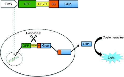
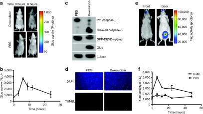
Noninvasive detection and in vivo imaging of apoptosis plays a critical role in the development of therapeutics in many different fields including cancer. We have developed an apoptosis biosensor by fusing green fluorescent protein (GFP) to the N-terminus of the naturally secreted Gaussia luciferase separated by a caspase-3 cleavage peptide consisting of aspartic acid (D), glutamic acid (E), valine (V), and aspartic acid (D) or DEVD. We showed that this fusion is retained in the cytoplasm of cells in an inactive form. Upon apoptosis, the DEVD peptide is cleaved in response to caspase-3 activation, freeing ssGluc, which can now enter the secretory pathway where it is folded properly and is released from the cells and can be detected in the conditioned medium in culture or in blood of live animals ex vivo over time. Because Gluc is secreted from cells via conventional pathway through the endoplasmic reticulum (ER), Golgi and vesicles, we showed that the presence of Gluc in these compartments in response to apoptosis can be visualized in vivo using bioluminescence imaging. This reporter provides a valuable tool for imaging and real-time monitoring of apoptosis and is compatible with high-throughput functional screening application in cultured cells and animal models.


Figures

Figure 1
Figure 1
Apoptosis biosensor. Schematic overview of the GFP-DEVD-ssGluc system. ptGFP lacking stop codon is fused to the N-terminal region of the Gaussia luciferase (including its signal sequence, ssGluc) separated by a DEVD, the caspase-3 cleavage peptide (GFP-DEVD-ssGluc). This fusion protein resides in the cytoplasm. Upon caspase-3 activation during apoptosis, the DEVD sequence is cleaved, freeing ssGluc, which can then enter the endoplasmic reticulum (ER) and be secreted into the conditioned medium of cultured cells or into blood of mice bearing cells expressing it in vivo. GFP, green fluorescent protein; ptGFP, Ptilosarcus GFP.
Figure 2
Figure 2
Real-time monitoring of apoptosis in culture. (a) Gluc is inactive when it resides in the cytoplasm. 293T human fibroblast cells were transfected in triplicates with plasmid expressing either native secreted Gluc (ssGluc), GFP-DEVD-ssGluc, or Gluc without the signal sequence (no ss). Forty-eight hours later, cell lysates or conditioned medium were assayed in triplicates for the Gluc activity. (b,c) Gli36 human glioma cells stably expressing GFP-DEVD-ssGluc were plated in a 6-well plate and treated with different amounts of doxorubicin in triplicates. (b) Twenty-four hours later, or at (c) different time points, 50 µl aliquots of the conditioned medium was collected in triplicates and assayed for Gluc activity using a luminometer. (d) Aliquots (in duplicates) of conditioned medium from (c) were imaged for Gluc activity using a CCD camera. For Gluc activity assay, data presented as mean ± SD for the three biological and three experimental replicates. GFP, green fluorescent protein.
Figure 3
Figure 3
GFP-DEVD-ssGluc is cleaved specifically in response to apoptosis. (a,b) Gli36 cells-expressing GFP-DEVD-ssGluc were treated with different concentration of doxorubicin in triplicates. Sixteen hours later, cell lysates as well as conditioned medium were analyzed by western blotting using an antibody against (a) Gluc and 50 µl aliquots of conditioned medium were assayed for (b) Gluc activity. (c,d) Gli36 cells-expressing GFP-DEVD-ssGluc were treated with either different amounts of doxorubicin in triplicates, or a combination of both doxorubicin and ZVAD-fmk, a caspase inhibitor. Sixteen hours later, cell lysates and conditioned medium were analyzed by western blotting for Gluc as well as caspase-3 using (c) specific antibodies and conditioned medium was assayed for (d) Gluc activity. Western blots shown are representative from a single well of different groups. For Gluc assay, data presented as mean ± SD for three biological and three experimental replicates.
Figure 4
Figure 4
Monitoring of extrinsic apoptosis and validation of Gluc secretion. (a) GFP-DEVD-ssGluc as a biosensor for extrinsic apoptosis. Gli36 cells-expressing GFP-DEVD-ssGluc were plated in a 96-well plate and treated with different concentrations of tumor necrosis factor-related apoptosis-inducing ligand (TRAIL) in triplicates. Sixteen hours later, aliquots of conditioned medium (in triplicates) were assayed for Gluc activity. (bc) Cleaved ssGluc is secreted from cells via conventional secretory pathway. Gli36 cells-expressing GFP-DEVD-ssGluc or native Gluc were treated with phosphate-buffered saline (PBS) or brefeldin A (BFA; 5 µg/ml) for 2 hours. Cells were then treated with either PBS or doxorubicin (0.8 µmol/l) in triplicates and, 8 hours later, conditioned media and lysates were assayed for Gluc activity. Data presented as mean ± SD for relative Gluc activity in which the signal obtained from control native ssGluc treated with PBS only in b or treated with dox only (no BFA) in c is set to 1.
Figure 5
Figure 5
In vivo imaging and ex vivo monitoring of apoptosis. (a) Gli36 cells-expressing GFP-DEVD-ssGluc were implanted subcutaneously in 10 nude mice. Two weeks later, tumors were injected with either PBS (control) or 10 µg of doxorubicin (n = 5 per group). Immediately before and after 8 hours injection, mice were intravenously (i.v.) injected with coelenterazine and imaged using a CCD camera. (b) In the same experiment, 5 µl blood (in triplicates) was withdrawn at different time points and assayed for Gluc activity using a luminometer. (c) A set of tumors from each group in a were removed 8 hours post-treatment and assayed by western blotting using antibodies against Gluc and caspase-3. (d) A set of mice from (a) were perfused with 4% paraformaldehyde in PBS, tumors were removed, fresh frozen, sectioned into 10-µm sections and analyzed using TUNEL (red) as well as DAPI (blue) staining. Bar = 100 µm. (e,f) MDA-MB-231BR breast cancer cells were injected into the left ventricle of female nude mice. Four weeks later, mice were imaged with Fluc bioluminescence after intraperitoneal (i.p.) injection of -luciferin. A representative mouse imaged front and back showing that these cells did metastasize (e). Mice were then i.p. injected with either PBS or TRAIL (n = 6 per group) and at different time points, 5 µl blood (in triplicates) was withdrawn at different time points and assayed for Gluc activity using a luminometer (f). Data shown as mean ± SD.

References

    1. Budihardjo I, Oliver H, Lutter M, Luo X., and, Wang X. Biochemical pathways of caspase activation during apoptosis. Annu Rev Cell Dev Biol. 1999;15:269–290. - PubMed
    1. Fesik SW. Promoting apoptosis as a strategy for cancer drug discovery. Nat Rev Cancer. 2005;5:876–885. - PubMed
    1. Moffitt KL, Martin SL., and, Walker B. Proteases implicated in apoptosis: old and new. J Pharm Pharmacol. 2010;62:563–576. - PubMed
    1. Blankenberg FG. Apoptosis imaging: anti-cancer agents in medicinal chemistry. Anticancer Agents Med Chem. 2009;9:944–951. - PubMed
    1. Fulda S., and, Debatin KM. Signaling through death receptors in cancer therapy. Curr Opin Pharmacol. 2004;4:327–332. - PubMed

Publication types