Review
. 2011 Feb;31(1):11-32.
doi: 10.1055/s-0031-1272832. Epub 2011 Feb 22.

Epithelial-mesenchymal interactions in biliary diseases

Affiliations
Review

Epithelial-mesenchymal interactions in biliary diseases

Luca Fabris et al. Semin Liver Dis. 2011 Feb.

Abstract

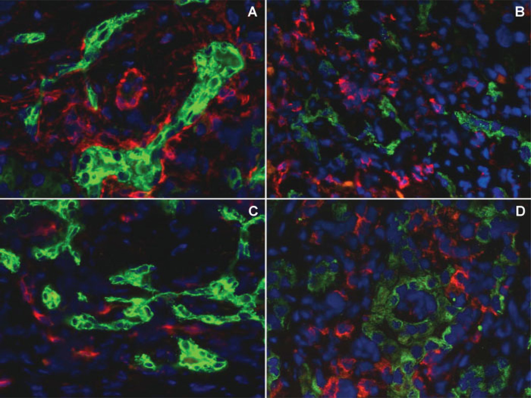
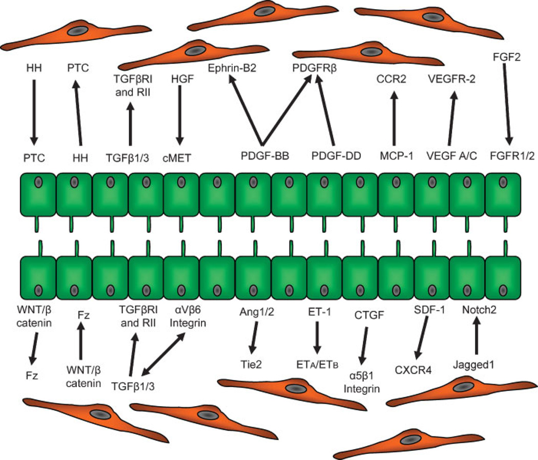
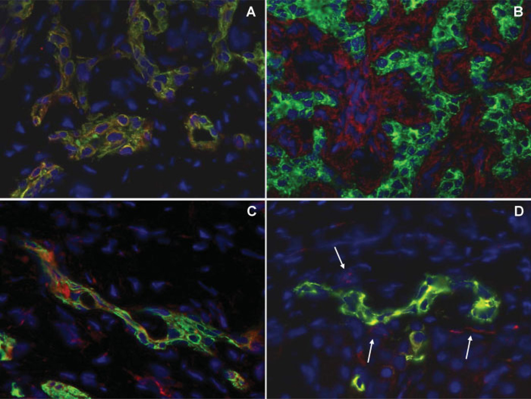
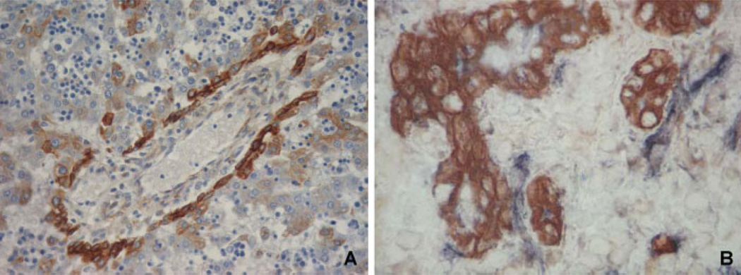
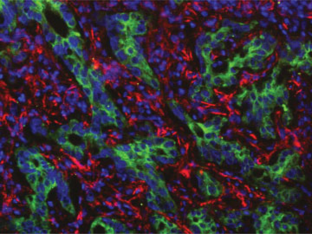
In most cholangiopathies, liver diseases of different etiologies in which the biliary epithelium is the primary target in the pathogenic sequence, the central mechanism involves inflammation. Inflammation, characterized by pleomorphic peribiliary infiltrate containing fibroblasts, macrophages, lymphocytes, as well as endothelial cells and pericytes, is associated to the emergence of "reactive cholangiocytes." These biliary cells do not possess bile secretory functions, are in contiguity with terminal cholangioles, and are of a less-differentiated phenotype. They have acquired several mesenchymal properties, including motility and ability to secrete a vast number of proinflammatory chemo/cytokines and growth factors along with de novo expression of a rich receptor machinery. These functional properties enable reactive cholangiocytes to establish intimate contacts and to mutually exchange a variety of paracrine signals with the different mesenchymal cell types populating the portal infiltrate. The extensive crosstalk between the epithelial and mesenchymal compartments is the driver of liver repair mechanisms in cholangiopathies, ultimately evolving toward portal fibrosis. Herein, the authors first review the properties of the different cell types involved in their interaction, and then analyze the underlying molecular mechanisms as they relate to liver repair in cholangiopathies.


Figures

Figure 1
Figure 1
Microanatomic relationships between reactive cholangiocytes and mesenchymal cell types in cholangiopathies. Dual-immunofluorescence of the biliary cell marker K19 (green fluorescence) with the myofibroblast marker α-SMA (red fluorescence, sample of biliary atresia, A), with the leucocyte marker CD45 (red fluorescence, biliary atresia, B), with the endothelial cell marker CD34 (red fluorescence, biliary atresia, C), and with the macrophage marker CD68 (red fluorescence, primary sclerosing cholangitis, D) shows that reactive cholangiocytes establish intimate contacts with multiple mesenchymal cell types. This represents the structural basis to mutually exchange a huge variety of paracrine signals. (Original magnification A–D: ×400)
Figure 2
Figure 2
Functional elements of the crosstalk between reactive cholangiocytes and hepatic stellate cells/myofibroblasts. A high homology of agonists/receptor systems is shared by reactive cholangiocytes and myofibroblasts that enable them to an extensive crosstalk, the molecular mechanism responsible for portal fibrogenesis. Biologic significance of the different systems involved in the crosstalk is summarized in Table 1.
Figure 3
Figure 3
Platelet-derived growth factor (PDGF) and vascular endothelial growth factor (VEGF) are strongly expressed by reactive cholangiocytes and mediate paracrine communications with mesenchymal cells. Dual immunofluorescence of the biliary cell marker K19 (green fluorescence) with PDGF-B (red fluorescence, A) and with PDGFR-β (red fluorescence, B) shows strong expression of PDGF-B on reactive cholangiocytes (coincident staining, yellow fluorescence), while its cognate receptor PDGFR-β extensively decorates multiple mesenchymal cells in the portal tract, in proximity to K19-positive ductular structures. Reactive cholangiocytes stained by K19 (green fluorescence) also co-express VEGF (coincident staining, yellow fluorescence, C), that signals to VEGFR-2 expressed by fibroblasts and endothelial cells adjacent to reactive ductules (paracrine loop, arrows) and also by reactive cholangiocytes themselves (autocrine loop) (single staining, red fluorescence; coincident staining, yellow fluorescence, D). These findings are consistent with a central role played by PDGF and VEGF in biliary repair, as seen in these tissue samples derived from a patient with biliary atresia undergoing liver transplantation. (Original magnification A-D: ×400)
Figure 4
Figure 4
SDF-1/CXCR4 crosstalk in biliary repair. Immunohistochemistry for SDF-1 shows its strong expression by ductal plate cells, starting from the earliest maturation ages when ductal plates are still organized as single layer cord, as seen in this sample obtained from abortive material at 16-gestation weeks (A, Original magnification ×200). Dual immunoperoxidase staining for SDF-1 (brown color) and CXCR4 (blue) in a tissue sample obtained from a patient with drug cholestatic injury, shows their expression is finely balanced in the epithelial and mesenchymal compartment (B, Original magnification ×400). Whereas SDF-1 is expressed by reactive cholangiocytes, CXCR4 is expressed by strictly adjacent mesenchymal cells. Given the brisk ductular expansion following biliary damage, reactive cholangiocyte-derived SDF-1 greatly increases and therefore facilitates recruitment and homing of CXCR4-positive cells, among which are fibrocytes.
Figure 5
Figure 5
Abundant CAF enrichment in cholangiocarcinoma. Dual immunofluorescence for the cholangiocyte marker K19 (green fluorescence) and the CAF marker α-SMA (red fluorescence) in a tumoral sample of cholangiocarcinoma obtained from surgical resection. Extensive recruitment of CAF is observed among neoplastic bile ducts, leading to the formation of a rich tumor reactive stroma that represents a distinctive feature of CCA. This feature is relevant for cancer growth, given the ability of CAF to provide cancer cells with proliferative and antiapoptotic signals. (Original magnification ×200)

References

    1. Lazaridis KN, Strazzabosco M, Larusso NF. The cholangiopathies: disorders of biliary epithelia. Gastroenterology. 2004;127(5):1565–1577. - PubMed
    1. Strazzabosco M, Fabris L, Spirli C. Pathophysiology of cholangiopathies. J Clin Gastroenterol. 2005;39(4) Suppl 2:S90–S102. - PubMed
    1. Spirlì C, Fabris L, Duner E, et al. Cytokine-stimulated nitric oxide production inhibits adenylyl cyclase and cAMPdependent secretion in cholangiocytes. Gastroenterology. 2003;124(3):737–753. - PubMed
    1. Liu Z, Sakamoto T, Ezure T, et al. Interleukin-6, hepatocyte growth factor, and their receptors in biliary epithelial cells during a type I ductular reaction in mice: interactions between the periductal inflammatory and stromal cells and the biliary epithelium. Hepatology. 1998;28(5):1260–1268. - PubMed
    1. Terada R, Yamamoto K, Hakoda T, et al. Stromal cell-derived factor-1 from biliary epithelial cells recruits CXCR4-positive cells: implications for inflammatory liver diseases. Lab Invest. 2003;83(5):665–672. - PubMed

Publication types