. 2011 Apr 15;519(6):1007-23.
doi: 10.1002/cne.22570.

Quantitative study of NPY-expressing GABAergic neurons and axons in rat spinal dorsal horn

Affiliations
Free PMC article

Quantitative study of NPY-expressing GABAergic neurons and axons in rat spinal dorsal horn

Erika Polgár et al. J Comp Neurol. .
Free PMC article

Abstract

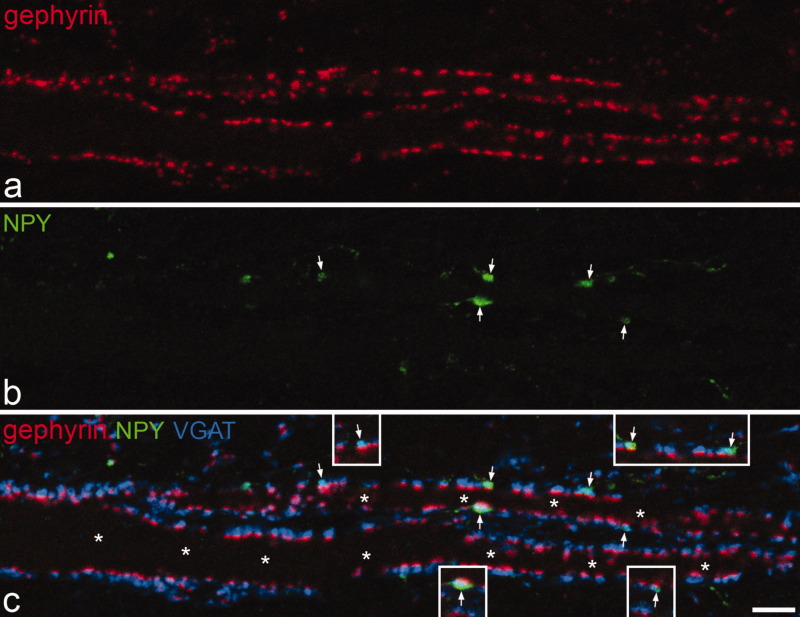
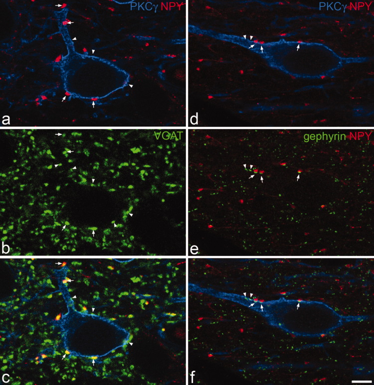
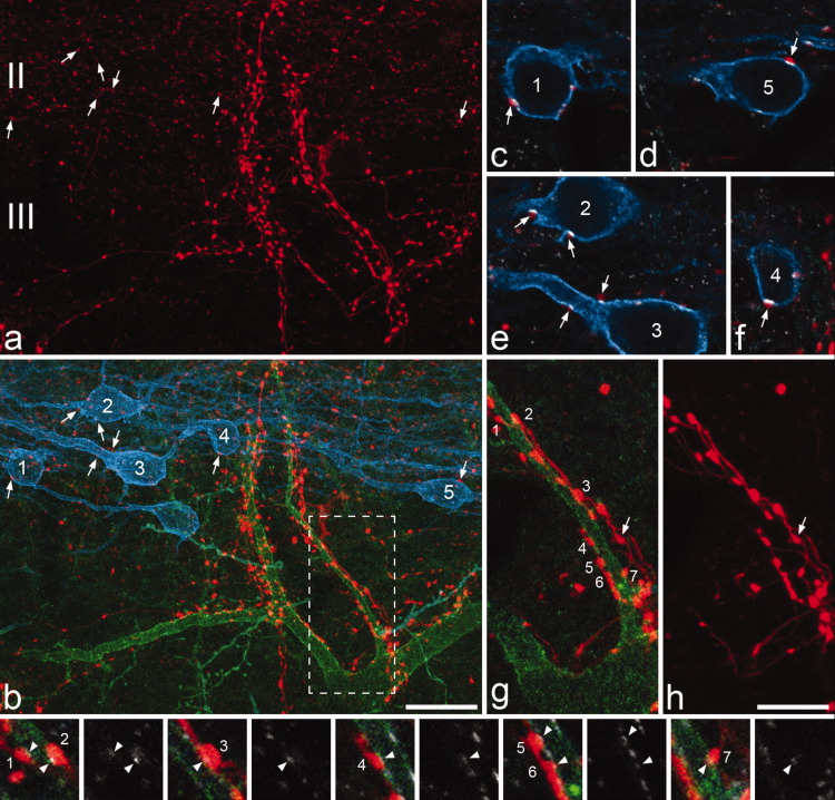
Between 25-40% of neurons in laminae I-III are GABAergic, and some of these express neuropeptide Y (NPY). We previously reported that NPY-immunoreactive axons form numerous synapses on lamina III projection neurons that possess the neurokinin 1 receptor (NK1r). The aims of this study were to determine the proportion of neurons and GABAergic boutons in this region that contain NPY, and to look for evidence that they selectively innervate different neuronal populations. We found that 4-6% of neurons in laminae I-III were NPY-immunoreactive and based on the proportions of neurons that are GABAergic, we estimate that NPY is expressed by 18% of inhibitory interneurons in laminae I-II and 9% of those in lamina III. GABAergic boutons were identified by the presence of the vesicular GABA transporter (VGAT) and NPY was found in 13-15% of VGAT-immunoreactive boutons in laminae I-II, and 5% of those in lamina III. For both the lamina III NK1r-immunoreactive projection neurons and protein kinase Cγ (PKCγ)-immunoreactive interneurons in lamina II, we found that around one-third of the VGAT boutons that contacted them were NPY-immunoreactive. However, based on differences in the sizes of these boutons and the strength of their NPY-immunoreactivity, we conclude that these originate from different populations of interneurons. Only 6% of VGAT boutons presynaptic to large lamina I projection neurons that lacked NK1rs contained NPY. These results show that NPY-containing neurons make up a considerable proportion of the inhibitory interneurons in laminae I-III, and that their axons preferentially target certain classes of dorsal horn neuron.


Figures

Figure 1
Figure 1
The dorsal horn seen with darkfield illumination. The two rows of arrowheads show the dorsal and ventral borders of lamina II, which appears as a dark band. Scale bar = 100 μm.
Figure 2
Figure 2
NPY-immunostaining in the dorsal horn. A confocal image from a transverse section through the dorsal horn immunostained to reveal NPY. The dashed lines represent the ventral borders of laminae I, II, and III. There is a dense plexus of immunoreactive axons that occupies laminae I and II, with some axonal staining in deeper laminae. Dorsoventrally orientated bundles of axons can often be seen and two of these are marked with arrows. Scattered immunoreactive cell bodies are present throughout laminae I–III and some of these are indicated with arrowheads. Note that many of those in laminae I–II are hidden by the axonal plexus. The image is a projection of 24 optical sections at 1-μm z-spacing. Scale bar = 100 μm.
Figure 3
Figure 3
Confocal images showing NPY-immunoreactive neurons in laminae I–II. a: Part of a transverse section of the spinal cord immunostained to reveal NPY (green). b: The same field scanned for NeuN (blue) and the nuclear stain propidium iodide (red). In the merged image (c) the solid line indicates the edge of the gray matter, while the dashed line represents the lamina I/II border. Two NPY-immunoreactive cells (arrows) can be seen in lamina II. In each case the cell is NeuN-positive. Many other neurons that are not NPY-immunoreactive are visible. Nonneuronal nuclei lack NeuN, and therefore appear red. The small green profiles correspond to NPY-immunoreactive axons and their varicosities. The images were obtained from a single optical section. Scale bar = 10 μm.
Figure 4
Figure 4
The distribution of NPY-immunoreactive and nonimmunoreactive neurons in laminae I–III. A plot of all of the neurons included in the disector sample from one of the sections that was used to determine the proportion of neurons that were NPY-immunoreactive. NPY-positive cells are shown as filled circles, while the remaining neurons are shown as open circles.
Figure 5
Figure 5
NPY and VGAT in the superficial dorsal horn. a–c: Confocal images of a single optical section showing part of lamina I in a transverse section that had been reacted to reveal NPY (green) and VGAT (magenta). Several NPY-immunoreactive axonal boutons are visible, and some of these are indicated with arrows. As seen in the merged image (c), all of these boutons are also VGAT-immunoreactive, but they are surrounded by many other VGAT-positive boutons that lack NPY. The very fine green profiles (three indicated with arrowheads) were found to be intervaricose portions of NPY-immunoreactive axons when followed through the series of optical sections, and these did not contain VGAT. d: A projection of 24 optical sections at 0.3 μm z-spacing from the same field shows the distinction between boutons (arrows) and intervaricose portions (arrowheads) of NPY-immunoreactive axons. Scale bar = 5 μm.
Figure 6
Figure 6
Contacts between NPY-immunoreactive boutons and a lamina III NK1r-expressing neuron. a: A confocal image from lamina II in a parasagittal section that shows NK1r-immunoreactivity (blue) on part of the dorsal dendrite of a lamina III neuron. The dendrite receives several contacts from boutons that are NPY-immunoreactive (red), some of which are indicated with arrows. b: The same field scanned to reveal VGAT (green). c: The merged image reveals that many VGAT-immunoreactive boutons that lack NPY are also in contact with the dendrite (some shown with arrowheads). The images are from a single optical section. Scale bar = 5 μm.
Figure 7
Figure 7
Contacts between NPY-containing boutons and a large gephyrin-coated lamina I neuron. a: Immunostaining for gephyrin (red, TSA method) in a horizontal section of lamina I shows parts of two proximal dendrites that belong to a large neuron with a cell body in lamina I. b: The same field scanned to reveal NPY (green). c: Merging the staining for gephyrin, NPY, and VGAT (blue) reveals that virtually every gephyrin punctum on the surface of the cell is associated with a VGAT-positive bouton, and that a few of these (marked with arrows) are NPY-immunoreactive. Asterisks in c indicate the two dendritic shafts. Note that the gephyrin puncta are relatively large (compared to those illustrated in Fig. 8) because they were detected with the TSA method. The images are projections of three optical sections at 0.5 μm z-spacing. The insets in c show single optical sections through each of the contacts from NPY-containing boutons. Scale bar = 5 μm.
Figure 8
Figure 8
Contacts between NPY-containing boutons and PKCγ-immunoreactive interneurons in lamina IIi. a–c: show a field scanned to reveal PKCγ (blue), NPY (red), and VGAT (green) from a parasagittal section. A PKCγ-immunoreactive cell receives contacts on its cell body and dendrite from several VGAT-positive boutons, some of which also contain NPY. Arrows indicate some of the NPY boutons in contact with the cell and arrowheads show examples of VGAT boutons that do not contain NPY. d–f: A PKCγ cell (blue) in a section reacted to reveal NPY (red) and gephyrin (green). Arrows indicate three contacts onto the cell from NPY boutons and a gephyrin punctum is present in the cell membrane at each of these. Arrowheads show the locations of other gephyrin puncta that are in the cell membrane, but are not associated with NPY boutons. All images are from single optical sections. Scale bar = 5 μm.
Figure 9
Figure 9
NPY axons that innervate NK1r lamina III cells and PKCγ lamina II interneurons appear to originate from different sources. a,b: Part of laminae II and III in a sagittal section that had been immunostained for NPY (red), NK1r (green), and PKCγ (blue). Several PKCγ-immunoreactive neurons can be seen and five of these are numbered. Each of these cells has contacts from NPY-immunoreactive boutons. Some of these are marked with arrows and shown at higher magnification in c–f. A large NK1r-immunoreactive cell is also present in this field and this has numerous contacts from NPY boutons. c–f: Single confocal optical sections scanned to reveal NPY (red), PKCγ (blue), and gephyrin (white) show that a gephyrin punctum is present at each of the contacts that the PKCγ cells receive from NPY boutons (arrows). g,h: Show contacts from NPY-containing boutons onto one of the dorsal dendrites of the NK1r cell at higher magnification and correspond to the boxed region in b. Seven of the NPY boutons that contact the cell are identified with numbers. The images in the bottom row, each of which is from a single optical section, are in pairs. In each case the left one was scanned for NPY (red), NK1r (green), and gephyrin (white), while the right one shows only gephyrin. Each of the contacts between the NPY boutons and the dendrite of the NK1r cell is associated with a gephyrin punctum, but these are much paler than those on the PKCγ cells. Note in h that the NPY-immunoreactive boutons that contact the NK1r cell are often linked by clear intervaricose portions, which indicates that they must originate from the same neuron. However, these axons also give rise to boutons that are not in contact with the NK1r cell (an example is indicated with an arrow in g,h). The NPY boutons that are presynaptic to the PKCγ cells are generally paler (as shown in a), and were never found to be connected by visible intervaricose portions to those that were presynaptic to the NK1r cells. a,b: Projections of 34 optical sections and g,h: of 8 optical sections at 0.5 μm z-spacing. Scale bars = 20 μm in a,b; 10 μm in c–h.
Figure 10
Figure 10
Differences between NPY boutons that were presynaptic to lamina III NK1r projection neurons and PKCγ interneurons in lamina IIi. Frequency histograms show (a) the cross-sectional area and (b) normalized mean luminance values for boutons that were presynaptic to the lamina III NK1r projection neurons (black bars, n = 309) or the PKCγ interneurons (gray bars, n = 280). In both cases the differences between these two populations were highly significant (P < 0.0001, Mann–Whitney U-test).

Similar articles

Cited by

References

    1. Al-Khater KM, Kerr R, Todd AJ. A quantitative study of spinothalamic neurons in laminae I, III, and IV in lumbar and cervical segments of the rat spinal cord. J Comp Neurol. 2008;511:1–18. - PMC - PubMed
    1. Antal M, Polgár E, Chalmers J, Minson JB, Llewellyn-Smith I, Heizmann CW, Somogyi P. Different populations of parvalbumin- and calbindin-D28k-immunoreactive neurons contain GABA and accumulate 3H-D-aspartate in the dorsal horn of the rat spinal cord. J Comp Neurol. 1991;314:114–124. - PubMed
    1. Antal M, Petkó M, Polgár E, Heizmann CW, Storm-Mathisen J. Direct evidence of an extensive GABAergic innervation of the spinal dorsal horn by fibres descending from the rostral ventromedial medulla. Neuroscience. 1996;73:509–18. - PubMed
    1. Becker CM, Hoch W, Betz H. Sensitive immunoassay shows selective association of peripheral and integral membrane proteins of the inhibitory glycine receptor complex. J Neurochem. 1989;53:124–131. - PubMed
    1. Bjugn R, Gundersen HJ. Estimate of the total number of neurons and glial and endothelial cells in the rat spinal cord by means of the optical disector. J Comp Neurol. 1993;328:406–414. - PubMed

Publication types

MeSH terms

LinkOut - more resources