. 2011 Apr 15;519(6):1165-80.
doi: 10.1002/cne.22566.

Cytoarchitecture of the lateral ganglionic eminence and rostral extension of the lateral ventricle in the human fetal brain

Affiliations

Cytoarchitecture of the lateral ganglionic eminence and rostral extension of the lateral ventricle in the human fetal brain

Hugo Guerrero-Cázares et al. J Comp Neurol. .

Abstract

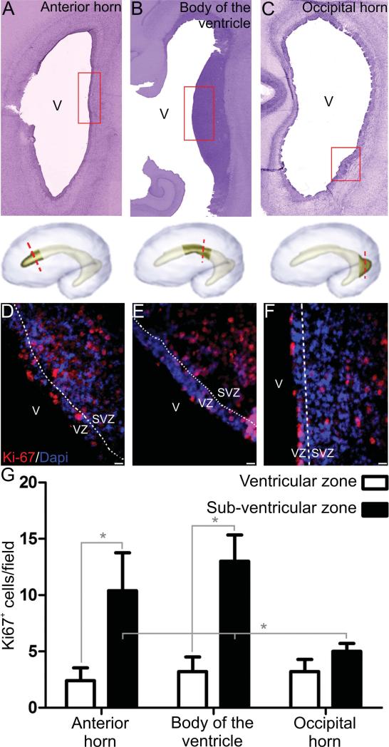
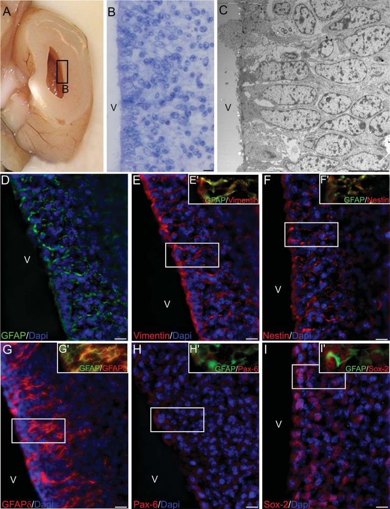
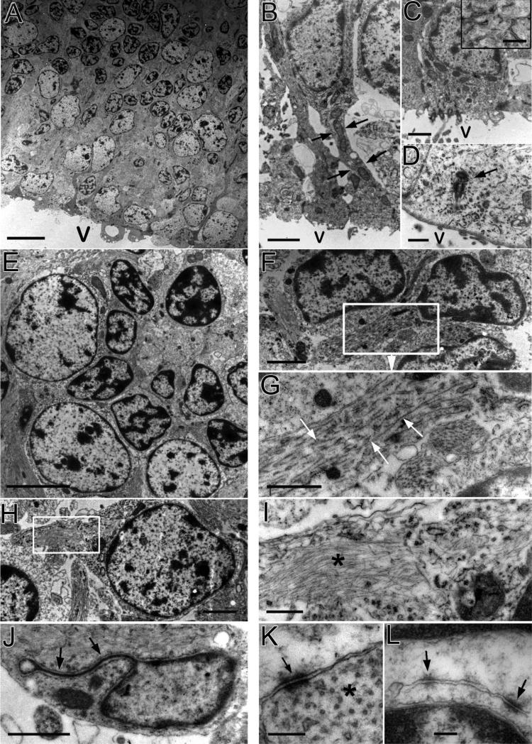
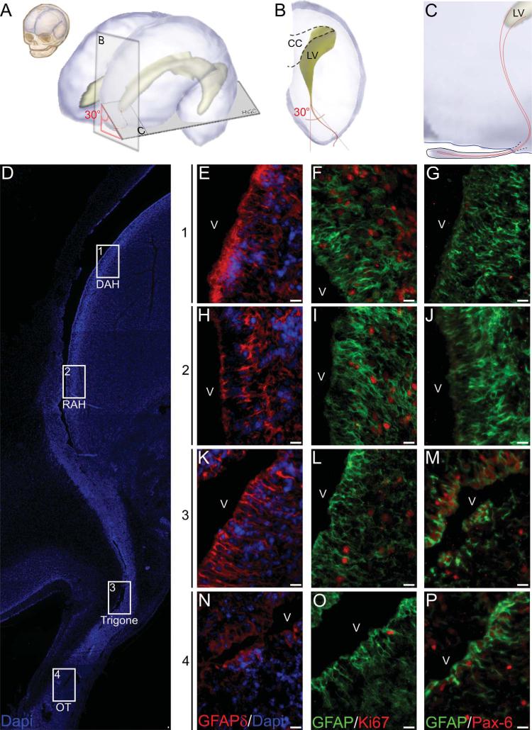
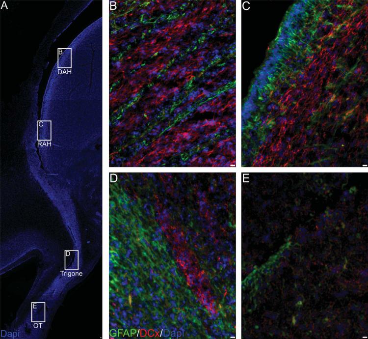
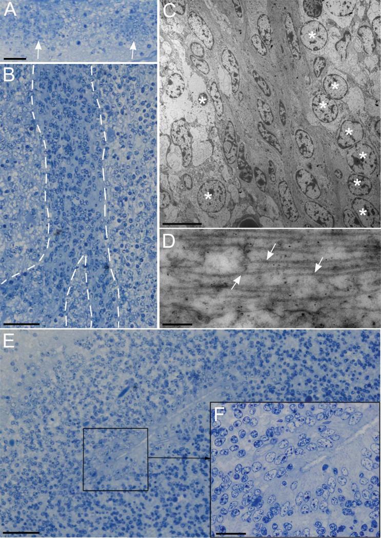
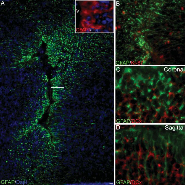
The fetal development of the anterior subventricular zone (SVZ) involves the transformation of radial glia into neural stem cells, in addition to the migration of neuroblasts from the SVZ towards different regions in the brain. In adult rodents this migration from the anterior SVZ is restricted to the olfactory bulb following a rostral migratory stream (RMS) formed by chains of migratory neuroblasts. Similar to rodents, an RMS has been suggested in the adult human brain, where the SVZ remains as an active proliferative region. Nevertheless, a human fetal RMS has not been described and the presence of migratory neuroblasts in the adult remains controversial. Here we describe the cytoarchitecture of the human SVZ at the lateral ganglionic eminence late in the second trimester of development (23-24 weeks postconception). Cell organization in this region is heterogeneous along the ventricular wall, with GFAP-positive cells aligned to the ventricle. These cells coexpress markers for radial glia like GFAPδ, nestin, and vimentin. We also show the presence of abundant migratory neuroblasts in the anterior horn SVZ forming structures here denominated cell throngs. Interestingly, a ventral extension of the lateral ventricle suggests the presence of a putative RMS. Nevertheless, in the olfactory bulb neuroblast throngs or chain-like structures were not observed. The lack of these structures closer to the olfactory bulb could indicate a destination for the migratory neuroblasts outside the olfactory bulb in the human brain.


Figures

Figure 1
Figure 1
Cell proliferation in the ventricular zone (VZ) and subventricular zone (SVZ) of the lateral ganglionic eminence (LGE) and caudal ganglionic eminence. Representative images of coronal sections from the human fetal anterior horn (A), body of the ventricle (B), and occipital horn (C), stained with cresyl violet. Red rectangles show the areas where Ki67 expression was quantified. D–F: Representative images of Ki67 immunostains at different regions. G: Ki67+ cell quantification shows a higher proliferation rate in the SVZ when compared to the VZ. Higher proliferation is more evident in the anterior horn and the body of the ventricle when compared to the occipital horn *P < 0.05. V, Ventricle; VZ, ventricular zone; SVZ, subventricular zone. Scale bars = 10 μm.
Figure 2
Figure 2
Cell organization of the anterior horn (AH) LGE. A: Coronal section of a human fetal brain anterior horn. B: Cresyl violet-stained section showing a pseudostratified epithelium. C: EM image showing cells touching the ventricular wall, with cell nuclei arranged at different levels forming the pseudostratified appearance. D: Ventricular epithelial cells express GFAP filaments and arrange their process in a radial manner, these cell elongations coexpress vimentin (E), nestin (F), and GFAPδ (G). No specific expression of Pax-6 was observed in this region (H) while Sox-2 was abundant in both the VZ and the SVZ (I). Insets (E′–I′) show coexpression of the different proteins with GFAP. V, ventricle. Scale bars = 10 μm.
Figure 3
Figure 3
EM analysis of the AH LGE. A: Two cell types (large-clear and amall-dark) are observed in this region. B: A pseudostratified epithelium is formed by cell nuclei distributed adjacent to the ventricular wall or several microns away. B: Some cells extend long apical processes to contact the ventricular wall (arrows). C,D: Multiciliated and uniciliated cells (respectively) are observed in this region, arrow shows the basal corpuscle and centriole of a uniciliated cell. E: Two types of SVZ cells are observed, one with large and clear cytoplasm and nucleus, with noncondensed chromatin, and one with small and dark cytoplasm and nucleus with more condensed chromatin. F: A group of small cells with dark nuclei and small ribosome-rich cytoplasm, small endoplasmic reticulum cisternae and little dictyosomes. These cells present abundant microtubuli as shown in G (arrows). H: Cells with clear nuclei are characterized by the presence of abundant intermediate filaments, organelles, and few ribosomes and endoplasmic reticulum cisternae. I: Detail showing the intermediate filaments (asterisk). J: Clear nucleus cell with areas of nuclear fusion (arrows), tight junctions (arrows) can be observed between clear and dark nucleus cells (K); and between two clear cells (L). Asterisk in K shows transversal sections of intermediate filaments found in a dark nucleus cell. Scale bars = 10 μm in A; 2 μm in B,C,F,H (inset in C: 0.5 μm); 1 μm in D,G,I; E: 5 μm; 0.5 μm in J; 200 nm in K,L.
Figure 4
Figure 4
Rostral extension of the lateral ventricle in the human fetal brain. A: Diagram representing the human fetal brain at 20 weeks of gestation. B,C: The lateral ventricle anterior horn extends into the olfactory bulb at a lateral inclination of 30° from the ventricle. D: Semi-sagittal section, stained with the nuclear dye DAPI. The ventricular opening extends from the lateral ventricle to the olfactory bulb. Cells from the ventricular wall are positive for GFAPδ in all the regions studied (E,H,K,N). Ki-67-positive cells are more abundant in the DAH (F), RAH (I), and trigone (L) when compared to the OT (O). Pax-6-positive cells are absent in the DAH (G) and RAH (J) and present in the more rostral regions of the trigone (M) and the OT (P). CC, corpus callosum; LV lateral ventricle; V, ventricle; DAH, Dorsal anterior horn; RAH, Rostral anterior horn; OT, olfactory tract. Scale bars = 10 μm.
Figure 5
Figure 5
Doublecortin (DCx)-positive structures in the anterior SVZ. A: Reconstruction of the entire connection between the anterior horn and the olfactory tract. DCx+ cells were found at every region with different organization. At the DAH (B), RAH (C), and olfactory trigone (D) DCx+ cells were found aligned to GFAP-positive cells. At the OT, no GFAP+ cells were observed in alignment with the less abundant DCx+ cells. Scale bars = 10 μm.
Figure 6
Figure 6
A: Transversal section of the olfactory trigone where a large group of dark nucleus cells (arrows) is surrounded by clear nucleus cells. B: Longitudinal section of the olfactory tract showing a bifurcating chain of dark nucleus cells surrounded by clear nucleus cells. C: EM image of a chain of dark nucleus cells showing an elongated morphology. These cells are surrounded by groups of clear cells with round nuclei (*). D: High-magnification image showing microtubules in a dark nucleus cell. E,F: Semithin section of the olfactory bulb showing a ventricular lumen delimited by a pseudostratified epithelium. Scale bars = 50 μm in A,B,E; 8 μm in C; 250 nm in D; 20 μm in F.
Figure 7
Figure 7
Cytoarchitecture of the human fetal olfactory ventricle. A: GFAP-positive cells align to the olfactory bulb ventricle similarly to the anterior horn. These GFAP-positive cells also coexpress the GFAPδ isoform (inset). B: Ki67-positive cells are observed in the OB parenchyma but none were found in the ventricular wall. DCx-positive fibers are observed in this region but no clear alignment was observed either on coronal (C) or sagittal (D) sections. V, ventricle. Scale bars = 10 μm.

References

    1. Alonso G, Prieto M, Chauvet N. Tangential migration of young neurons arising from the subventricular zone of adult rats is impaired by surgical lesions passing through their natural migratory pathway. J Comp Neurol. 1999;405:508–528. - PubMed
    1. Alvarez-Buylla A, Kohwi M, Nguyen TM, Merkle FT. The heterogeneity of adult neural stem cells and the emerging complexity of their niche. Cold Spring Harb Symp Quant Biol. 2008;73:357–365. - PubMed
    1. Baer K, Eriksson PS, Faull RL, Rees MI, Curtis MA. Sox-2 is expressed by glial and progenitor cells and Pax-6 is expressed by neuroblasts in the human subventricular zone. Exp Neurol. 2007;204:828–831. - PubMed
    1. Bayer SA, Altman J. Atlas of human central nervous system development. CRC Press; Boca Raton, FL: 2007.
    1. Bedard A, Parent A. Evidence of newly generated neurons in the human olfactory bulb. Brain Res Dev Brain Res. 2004;151:159–168. - PubMed

Publication types