. 2011 Apr;20(4):641-55.
doi: 10.1002/pro.600. Epub 2011 Mar 7.

Structure and dynamics of cationic membrane peptides and proteins: insights from solid-state NMR

Affiliations

Structure and dynamics of cationic membrane peptides and proteins: insights from solid-state NMR

Mei Hong et al. Protein Sci. 2011 Apr.

Abstract

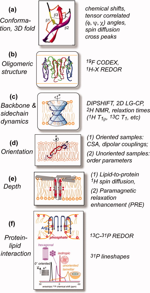
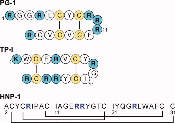
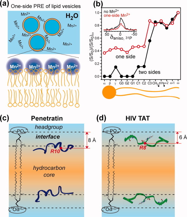
Many membrane peptides and protein domains contain functionally important cationic Arg and Lys residues, whose insertion into the hydrophobic interior of the lipid bilayer encounters significant energy barriers. To understand how these cationic molecules overcome the free energy barrier to insert into the lipid membrane, we have used solid-state NMR spectroscopy to determine the membrane-bound topology of these peptides. A versatile array of solid-state NMR experiments now readily yields the conformation, dynamics, orientation, depth of insertion, and site-specific protein-lipid interactions of these molecules. We summarize key findings of several Arg-rich membrane peptides, including β-sheet antimicrobial peptides, unstructured cell-penetrating peptides, and the voltage-sensing helix of voltage-gated potassium channels. Our results indicate the central role of guanidinium-phosphate and guanidinium-water interactions in dictating the structural topology of these cationic molecules in the lipid membrane, which in turn account for the mechanisms of this functionally diverse class of membrane peptides.


Figures

Figure 1
Figure 1
Summary of different types of membrane protein structural information that can be obtained from solid-state NMR. (a) Monomer conformation and 3D fold. (b) Oligomeric structure. (c) Protein dynamics. (d) Protein orientation in the membrane. (e) Depth of insertion. (f) Protein–lipid interactions.
Figure 2
Figure 2
Amino acid sequence of three disulfide-stabilized β-sheet AMPs.
Figure 3
Figure 3
The topology of Arg-rich membrane peptides reveals their mechanisms of action. The side views of the peptide structure and topology in the membrane are shown in the top row, whereas the bottom row shows either the peptide structure motif or the top view of the membrane-bound topology. (a) PG-1 forms TM β-barrels that cause toroidal pore defects in POPE/POPG membranes. Four to five PG-1 dimers comprise the barrel, which has an inner diameter of ∼20 Å. (b) TP-I binds to the interface of anionic lipid membranes and undergoes fast uniaxial diffusion to cause membrane defects. (c) HNP-1 dimers form TM pores where each dimer is oriented with the hydrophobic basket bottom facing the lipids and the hydrophilic top facing the pore. R25 is closest among the four Arg residues to the membrane surface. The monomer structure as determined by solid-state NMR is shown at the bottom. (d) The S4 helix of the KvAP voltage-sensing domain is inserted across the membrane with a 40° tilt angle and thins the membrane to stabilize the gating Arg molecules. The distribution of the Arg residues is shown in the helical wheel diagram at the bottom.
Figure 4
Figure 4
CPP-mediated intracellular delivery of therapeutic cargos.
Figure 5
Figure 5
Depth of insertion of CPPs. (a) One-side PRE of large unilamellar vesicles. Orange: lipids. Blue: water. Mn2+ ions are distributed only on the outer surface of the lipid vesicles. (b) Double normalized intensity of Mn2+-bound POPC/POPG membranes. 31P intensities are roughly halved in a one-side Mn2+ sample (inset) as expected. Carbons more embedded in the membrane experience less T2 relaxation enhancement and thus exhibit higher intensities. Red: one-side Mn2+-bound samples. Black: two-side Mn2+-bound samples. (c) Depth of penetratin in POPC/POPG membranes (P/L = 1:15) from one-side PRE and 13C-31P distances. (d) Depth of HIV TAT in DMPC/DMPG membranes (P/L = 1:15) from 1H spin diffusion and 13C-31P distances.
Figure 6
Figure 6
13C-31P distances and order parameters of several residues in membrane-bound penetratin (a–c) and TAT (d). (a) Arg10 Cζ-P distance in DMPC/DMPG-bound penetratin. (b) Lys13-phosphate distance in DMPC/DMPG bound penetratin. (c) Longer Ile3 13C-31P distances in penetratin indicate no interaction between neutral residues and lipid headgroups. (d) Guanidinium-phosphate and guanidinium-water interactions of Arg8 in DMPC/DMPG bound TAT. All distances were measured in gel-phase membranes while dipolar order parameters were measured in the liquid-crystalline phase.

Similar articles

Cited by

References

    1. Zasloff M. Antimicrobial peptides of multicellular organisms. Nature. 2002;415:389–395. - PubMed
    1. Fischer R, Fotin-Mleczek M, Hufnagel H, Brock R. Break on through to the other side-biophysics and cell biology shed light on cell-penetrating peptides. ChemBioChem. 2005;6:2126–2142. - PubMed
    1. Jiang Y, Lee A, Chen J, Ruta V, Cadene M, Chait BT, MacKinnon R. X-ray structure of a voltage-dependent K+ channel. Nature. 2003;423:33–41. - PubMed
    1. Hessa T, Kim H, Bihlmaier K, Lundin C, Boekel J, Andersson H, Nilsson I, White SH, von Heijne G. Recognition of transmembrane helices by the endoplasmic reticulum translocon. Nature. 2005;433:377–381. - PubMed
    1. Popot JL, Engelman DM. Membrane protein folding and oligomerization: the two-stage model. Biochemistry. 1990;29:4031–4037. - PubMed

MeSH terms

LinkOut - more resources