Review
. 2011 Feb 25;12(3):462-74.
doi: 10.1002/cphc.201000664. Epub 2010 Dec 29.

FRET microscopy in 2010: the legacy of Theodor Förster on the 100th anniversary of his birth

Affiliations
Review

FRET microscopy in 2010: the legacy of Theodor Förster on the 100th anniversary of his birth

Yuansheng Sun et al. Chemphyschem. .

Abstract

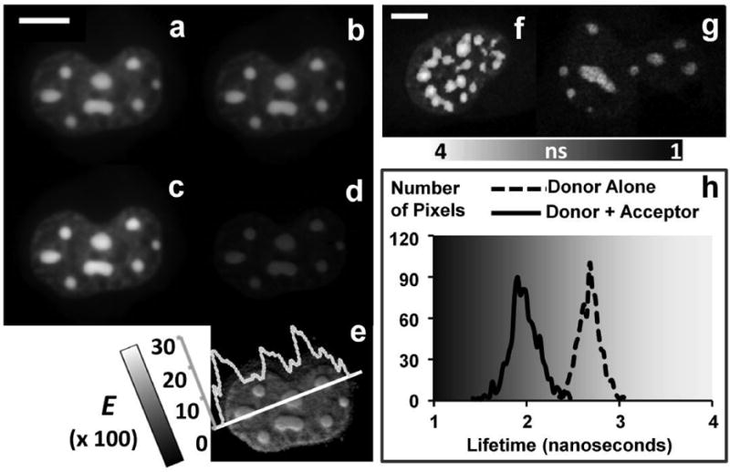
Theodor Förster would have been 100 years old this year, and he would have been astounded to see the impact of his scientific achievement, which is still evolving. Combining his quantitative approach of (Förster) resonance energy transfer (FRET) with state-of-the-art digital imaging techniques allows scientists to breach the resolution limits of light (ca. 200 nm) in light microscopy. The ability to deduce molecular or particle distances within a range of 1-10 nm in real time and to prove or disprove interactions between two or more components is of vital interest to researchers in many branches of science. While Förster's groundbreaking theory was published in the 1940s, the availability of suitable fluorophores, instruments, and analytical tools spawned numerous experiments in the last 20 years, as demonstrated by the exponential increase in publications. These cover basic investigation of cellular processes and the ability to investigate them when they go awry in pathological states, the dynamics involved in genetics, and following events in environmental sciences and methods in drug screening. This review covers the essentials of Theodor Förster's theory, describes the elements for successful implementation of FRET microscopy, the challenges and how to overcome them, and a leading-edge example of how Förster's scientific impact is still evolving in many directions. While this review cannot possibly do justice to the burgeoning field of FRET microscopy, a few interesting applications such as threecolor FRET, which greatly expands the opportunities for investigating interactions of cellular components compared with the traditional two-color method, are described, and an extensive list of references is provided for the interested reader to access.


Figures

Figure 1
Figure 1. Number of FRET publications by broad categories - Cumulative by year
(see resources at http://www.kcci.virginia.edu/Literature).
Figure 2
Figure 2. Basic concepts of FRET
FRET is the non-radiative energy transfer from an excited-state donor (D) to an acceptor (A) at the ground state, in close proximity (1∼10 nm), being a long-range dipole-dipole coupling mechanism. Thus, as illustrated in (a), FRET can be used to detect the interaction between fluorescently labeled cellular components within 1∼10 nm, far beyond the resolution limit of a light microscope (∼ 200 nm). Other than the D-A distance, FRET also requires two other conditions: (b) a significant overlap between the donor emission and the acceptor excitation spectra (covered by the grey area). (c) a favorable dipole moment - κ2 = (cosθT – 3 · cosθD · cosθA), where θT is the angle between the emission transition dipole of the donor and the absorption transition dipole of the acceptor; θD and θA are the angles between these dipoles and the vector joining the donor and the acceptor; κ2 cannot be 0 for FRET to occur and a larger κ2 increases the likelihood of FRET. (d) Since the energy transfer efficiency (E) from the donor to the acceptor is dependent on the inverse of the sixth power of the distance between them (see Equation 1), measuring E provides a sensitive indication on the D-A distance change around its Förster distance, as seen by plotting the relationship between the distance and the E of a single D-A pair, given its Förster distance of 5 nm to Equation 1.
Figure 3
Figure 3. Track the internalization of transferrin receptor-ligand complexes in live MDCK cells using confocal FRET microscopy
We assayed the distribution and sorting of receptor-ligand complexes in endocytic membranes, upon internalization from basolateral and/or apical plasma membrane domains [12, 13]. The ligands were conjugated to either Alexa488 (donor (D)) or Alexa555 (acceptor (A)) fluorophores. Confocal images of the double-labeled cells were obtained in the D (a), A (b) and FRET (c) channels. The processed FRET (PFRET, d) and apparent FRET efficiency (E%, e) images were obtained after processing the data with the PFRET algorithm in combination of the images acquired from the single-labeled specimens (see Section 4.1.2). To analyze whether these cellular trafficking steps occur in a random or clustered association between complexes, we made (f) a selection of regions of interest (ROIs) based on the uncorrected FRET image (c), and generated two parameters from the ROIs that determine a clustered distribution of complexes: (g) E%'s negative dependence on the D:A ratio and (h) E%'s independence of the acceptor level. The opposite is true for a random distribution, where E% is independent of the D:A ratio and dependent on the acceptor level. (Images were acquired by the Biorad Radiance 2100 confocal/multiphoton imaging system coupled to a Nikon TE 300 microscope equipped with a Nikon 60×/1.4NA water objective lens. The Argon 488 nm and HeNe 543 nm lasers were used as the donor and acceptor excitations, respectively. The 528/30 nm and 590/70 nm band-pass filters were used for the donor and acceptor emission channels, respectively. Scale bar = 10 μm.)
Figure 4
Figure 4. Demonstrate the homo-dimerization of CCAAT/enhancer-binding protein alpha (C/EBPα) in live mouse pituitary cell nucleus using both wide-field FRET microscopy and two-photon FLIM-FRET microscopy
Fluorescent signals from the nucleus of a cell co-expressing CFP-C/EBPα (donor (D)) and YFP-C/EBPα (acceptor (A)) were measured in the D (a), A (b) and FRET (c) channels in wide-field microscopy. The processed FRET (PFRET, d) and apparent FRET efficiency (E%, e) images were obtained after processing the data with the PFRET algorithm in combination of the images acquired from the single-label expressing cells (see Section 4.1.2). The interaction between CFP- and YFP-tagged C/EBPα is demonstrated by the E% images and indicates the homo-dimerization of C/EBPα in regions of centromeric heterochromatin of cell nucleus. The same biological system was also studied using a time correlated single photon counting (TCSPC) FLIM-FRET method, where the unquenched (τD = 2.68 ns) and quenched (τDA = 1.98 ns) donor lifetimes were determined from cells only expressing CFP--C/EBPα (f and the dash line in h) and cells that co-express CFP-C/EBPα and YFP-C/EBPα (g and the solid line in h). The FRET efficiency (E = 26.12%), determined from “1 - τDA / τD”, confirms the results obtained in the intensity-based wide-field FRET measurements. (Wide-field images were acquired by an Olympus IX70 microscope equipped with a 60X/1.2NA water objective lens and a CCD camera (Hamamatsu Orca2). The donor and acceptor excitation wavelengths were selected from the X-Cite ® 120 fluorescence illumination system (www.exfo-xcite.com) using 436/20 nm and 500/20 nm band-pass filters, respectively. The 470/30 nm and 535/30 nm band-pass filters were used to for the donor and acceptor emission channels, respectively. The TCSPC FLIM system and data analysis were described in detail earlier [65]. Data was acquired using the Becker & Hickl SPC-730 module on the Biorad Radiance 2100 confocal/multiphoton imaging system coupled to a Nikon TE 300 microscope equipped with a Nikon 60×/1.4NA water objective lens. Specimens were excited by a multi-photon laser tuned to 820 nm and the CFP signals were collected using a fast PMT (PMH-100, Becker & Hickl) with a 480/30 nm band-pass filter. Scale bar = 10 μm in both case.)
Figure 5
Figure 5. Comparison of FRET measurements of Cerulean (C)-Venus (V) based FRET-standard constructs (CTV, C32V, C17V, C5V) expressed in living cells in spectral FRET (sFRET), time correlated single photon counting (TCSPC) and frequency-domain (FD) FLIM-FRET microscopy (see Section 4.5)
(a) The average FRET efficiencies (E%) of the four FRET-standard constructs measured in all three methods are compared as columns with error bars being standard deviations (n = 12 for each construct measured in each method). (b) In sFRET microscopy, it is clearly observed from the raw spectral graphs that the ratio of the V peak to the C peak increases from CTV to C5V, indicating an increase in E% in the same direction. The quantitative confirmation is shown by the representative E% images obtained after processing the data with the sFRET algorithm (see Section 4.1.3). (c) shows the representative C-alone and FRET-standard decay profiles (at one pixel of each cell) and lifetime distributions (in each cell) obtained in the TCSCP FLIM-FRET measurements, clearly demonstrating a faster decay (a shorter lifetime) from C to C5V. (d) displays the phasor plots [97, 98] of the representative cells expressing C only or the FRET-standard constructs (20MHz, Semicircle - Single Lifetime Curve; (1,0) - Zero Lifetime; (0,0) - Infinite Lifetime), which also clearly indicate a longer lifetime from C5V to C. (The details about these measurements were described earlier [96]. Briefly, sFRET imaging was carried out on a Zeiss 510 Meta imaging system coupled to a Zeiss Axiovert 200M microscope equipped with a Zeiss 63X/1.4NA oil objective lens. The Argon 458 nm and 514 nm laser lines were used as the donor and acceptor excitations, respectively. The emission signals were acquired in the spectrum of 458∼651 nm at 10.7 nm intervals. The TCSPC FLIM data was acquired using the Becker Hickl SPC-150 module on the Biorad Radiance 2100 confocal/multiphoton imaging system coupled to a Nikon TE 300 microscope equipped with a Nikon 60×/1.4NA water objective lens. Specimens were excited by a multi-photon laser tuned to 820 nm and the Cerulean signals were collected using a fast PMT (PMC-100-0, Becker & Hickl) with a 480/30 nm band-pass filter. The FD FLIM measurements were carried on the ISS ALBA system coupled to a Nikon TiU microscope equipped with a Nikon 60×/1.4NA water objective lens. Specimens were excited by a 440 nm pulsed diode laser and the Cerulean signals were collected using an avalanche photodiode (APD) with a 480/30 nm band-pass filter. The phase shifts and amplitude attenuations were measured at five frequencies - 20, 40, 60, 80, 100 MHz.)
Figure 6
Figure 6. Three-color FRET model
(a) F1, F2, and F3 represent the excitation (dashed line) and emission (solid line) for the three fluorophores; the spectral overlap (donor emission and acceptor excitation) of each pair (F1-F2, F1-F3, F2-F3) is sufficient for FRET to occur. (b) Exciting the three-fluorophore system with Ex1 (solid lines), direct absorption and energy transfer occurs in parallel: F1→F2 at an energy transfer efficiency E12 and F1→F3 at an energy transfer efficiency E13; In addition, F2 (excited by Ex1 and sensitized due to the energy transfer F1→F2) also transfers energy to F3 at an energy transfer efficiency E23. At Ex2, the three-fluorophore system (dashed lines) becomes a 2-color (F2-F3) FRET system with F2 transferring energy to F3 at the same energy transfer efficiency E23. There is no energy transfer in the three-fluorophore system at Ex3 excitation. All of the above is based on the validated assumptions that the absorption rates of F1 at Ex2 or Ex3 and of F2 at Ex3 are trivial or not apparent.
Figure 7
Figure 7. Validation of the 3-color spectral FRET (3sFRET) microscopy method using FRET-standard constructs
To validate the 3sFRET method, three fluorescent proteins (FP) – mTFP, Venus and tdTomato (see Table 1) were used to build a 3-FP FRET-standard construct, where mTFP was coupled to Venus by a 5 amino acid (aa) linker and Venus was further tethered with tdTomato by a 10 aa linker, resulting in an mTFP-5aa-Venus-10aa-tdTomato complex. In addition, three 2-FP FRET-standard constructs were also generated: mTFP-5aa-Venus, Venus-10aa-tdTomato and mTFP-5aa-Amber-10aa-tdTomato, where Amber is a non-fluorescent mutant form of Venus (Y66C) [95], used in the 2-FP construct to maintain the same spatial relationship between mTFP and tdTomato as in the 3-FP construct. (a) is the overlay of the spectral images obtained from a cell expressing the 3-FP construct excited at the 458 nm wavelength and (b) shows the representative spectra of the cell sequentially excited at three different wavelengths, which were chosen around the peak excitation wavelengths of mTFP (458 nm), Venus (514 nm) and tdTomato (561 nm). The apparent FRET efficiencies (E%) between FPs in the 3-FP construct measured in 3sFRET microscopy - E12% (E% between mTFP and Venus, c), E13% (E% between mTFP and tdTomato, d) and E23% (E% between Venus and tdTomato, e), were validated by the E%s of the 2-FP constructs measured using an established 2-color spectral FRET microscopy method [50]: (f) mTFP-5aa-Venus, (g) mTFP-5aa-Venus-10aa- tdTomato and (h) Venus-5aa-tdTomato. (The details about the validation are described in Ref. [109]. Briefly, 3sFRET and two-color sFRET imaging was carried out on a Zeiss 510 Meta imaging system coupled to a Zeiss Axiovert 200M microscope equipped with a Zeiss 63X/1.4NA oil objective lens. The Argon 458 nm, 514 nm laser lines and diode-pumped solid-state 561 nm laser were used as the mTFP, Venus and tdTomato excitations, respectively. The emission signals were acquired in the spectrum of 469∼651 nm at 10.7 nm intervals. Scale bar = 10 μm.)
Figure 8
Figure 8. Demonstration of the homo-dimerization of CCAAT/enhancer-binding protein alpha (C/EBPα) and its interaction with heterochromatin protein 1 α (HP1α) in live mouse pituitary cell nucleus by 3-color confocal FRET Microscopy
Cells expressing mTFP- C/EBPα, Venus-C/EBPα and tdTomato-HP1α (see Table 1) were sequentially excited by three different wavelengths, which were chosen around the peak excitation (Ex) wavelengths of mTFP (Ex1), Venus (Ex2) and tdTomato (Ex3); the emitted signals were measured in three separate emission (Em) channels (mTFP: Em1, Venus: Em2, tdTomato: Em3), resulting the 6-channel images (a: Ex1-Em1, b: Ex1-Em2, c: Ex1-Em3, d: Ex2-Em2, e: Ex2-Em3 and f: Ex3-Em3). The images were then processed by the 3-color FRET algorithm, together with the images acquired from the single-label (mTFP-C/EBPα, Venus-C/EBPα and tdTomato-HP1α) control specimens in those 6 channels, to obtain the apparent FRET efficiency (E%) images (g: E% between mTFP and Venus, h: E% between mTFP and tdTomato, i: E% between Venus and tdTomato). The interactions between mTFP-C/EBPα, Venus-C/EBPα and tdTomato-HP1α are demonstrated by the 3-color E% images and indicate homo-dimerization of C/EBPα and the association between the C/EBPα dimer and HP1α in regions of heterochromatin of the cell nucleus. See Ref. [109] for more data analysis of this biological model in 3-color spectral FRET microscopy. (Confocal imaging was carried out on a Leica SP5 × white light laser (WLL) system [32] coupled to a Leica DMI6000 microscope equipped with a Leica 63×/1.4NA oil objective lens. The Argon 458 nm (Ex1), 514 nm (Ex2) and WLL 550 nm (Ex3) laser lines were used as the mTFP, Venus and tdTomato excitations, respectively. The mTFP (Em1 = 470∼500 nm), Venus (Em2 = 525∼550 nm) and tdTomato (Em3 = 560∼650 nm) emission channels were configured using the Leica acousto-optical beam splitter (AOBS). Scale bar = 10 μm.)

References

    1. Förster T. Naturwissenschafien. 1946;6:166–175.
    1. Clegg RM. In: Reviews in fluorescence. Geddes CD, Lakowicz JR, editors. Springer; New York: 2006. pp. 1–45.
    1. Gilmore AM, Larkum AW, Salih A, Itoh S, Shibata Y, Bena C, Yamasaki H, Papina M, Van Woesik R. Photochem Photobiol. 2003;77:515–523. - PubMed
    1. Förster T. In: modern quantum chemistry. Sinanoglu O, editor. Academic Press Inc.; 1965. pp. 93–137.
    1. Clegg RM. In: Fluorescence imaging spectroscopy and microscopy. Wang XF, Herman B, editors. John Wiley & Sons Inc.; New York: 1996. pp. 179–251.

MeSH terms

Personal name as subject

LinkOut - more resources