Review
. 2011 Feb 24:13:e6.
doi: 10.1017/S1462399410001754.

Emerging paramyxoviruses: molecular mechanisms and antiviral strategies

Affiliations
Review

Emerging paramyxoviruses: molecular mechanisms and antiviral strategies

Hector C Aguilar et al. Expert Rev Mol Med. .

Abstract

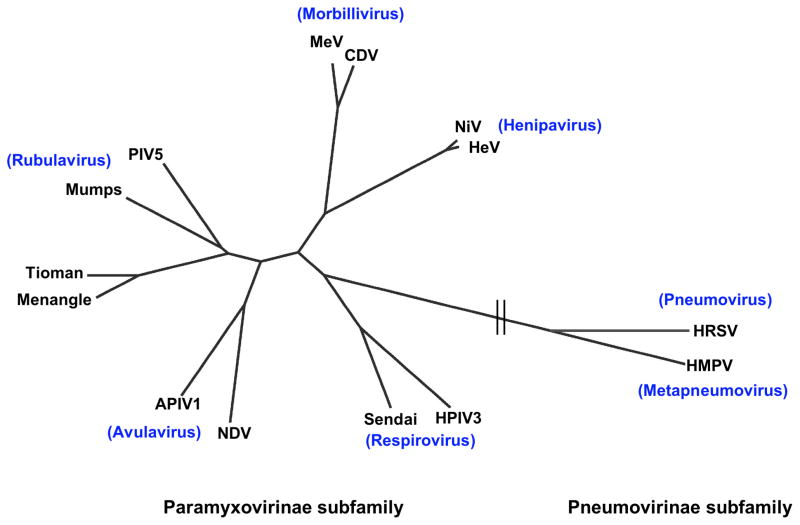
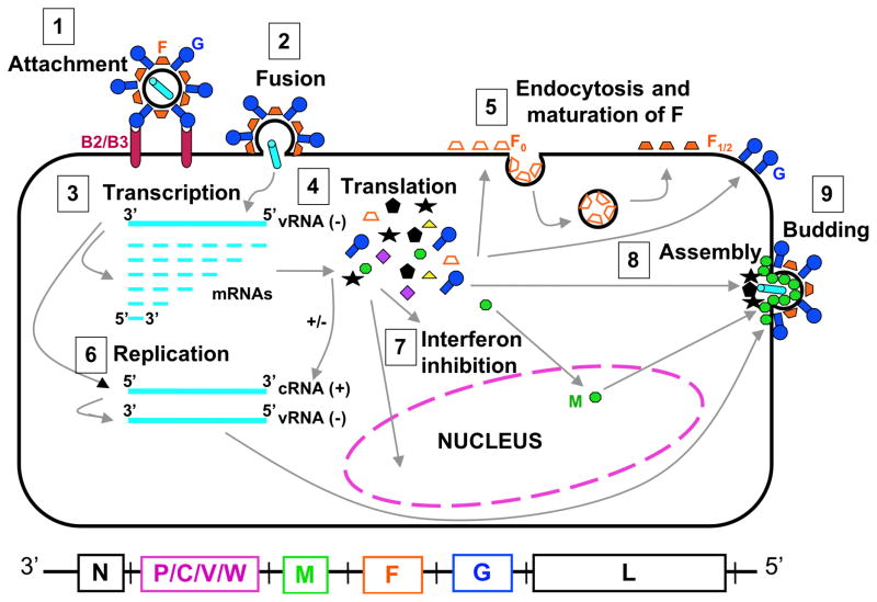
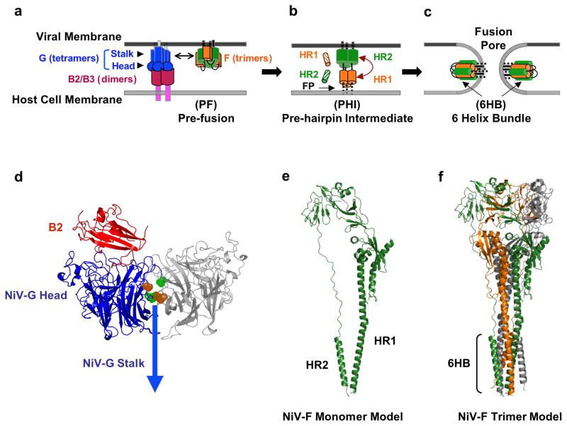
In recent years, several paramyxoviruses have emerged to infect humans, including previously unidentified zoonoses. Hendra and Nipah viruses (henipaviruses within this family) were first identified in the 1990s in Australia, Malaysia and Singapore, causing epidemics with high mortality and morbidity rates in affected animals and humans. Other paramyxoviruses, such as Menangle virus, Tioman virus, human metapneumovirus and avian paramyxovirus 1, which cause less morbidity in humans, have also been recently identified. Although the Paramyxoviridae family of viruses has been previously recognised as biomedically and veterinarily important, the recent emergence of these paramyxoviruses has focused our attention on this family. Antiviral drugs can be designed to target specific important determinants of the viral life cycle. Therefore, identifying and understanding the mechanistic underpinnings of viral entry, replication, assembly and budding will be critical in the development of antiviral therapeutic agents. This review focuses on the molecular mechanisms discovered and the antiviral strategies pursued in recent years for emerging paramyxoviruses, with particular emphasis on viral entry and exit mechanisms.


Figures

Fig. 1
Fig. 1. Phylogenetic tree of the Paramyxoviridae family, built using a fusion protein sequence comparison
The tree was generated from a Cobalt (NCBI) multiple fusion protein sequence alignment, by the fast minimum evolution method, and visualized using the Fig Tree program. Representative members of each genus of the Paramyxovirinae and Pneumovirinae subfamilies are shown. APIV1, avian parainfluenza virus 1; CDV, canine distemper virus; HeV, Hendra virus; HMPV, human metapneumovirus; HPIV3, human parainfluenza virus 3; HRSV, human respiratory syncytial virus; MeV, measles virus; NDV, Newcastle disease virus; NiV, Nipah virus; PIV-5, parainfluenza virus 5.
Fig. 2
Fig. 2. Henipavirus replication cycle
Depiction of henipavirus replication: After attachment to the B2/B3 receptor (1) and fusion (2), the virus enters the cell. The negative RNA genome (vRNA−) is a template for transcription of viral mRNAs following a 3–5′ attenuation gradient from N to L (3). mRNAs are translated into proteins (4) while the vRNA− is also a template for cRNA(+), which in turn is a template for vRNA(−) genomes during replication (6). New vRNA(−) genomes will be incorporated into new virions during viral assembly (8). Following translation (4), various viral proteins will function in interferon signaling pathways (7), and F0 will be endocytosed and matured (5). Assembly (8) and budding (9) are orchestrated primarily by the M protein, and N, P, C, M, F, and G, are incorporated into virions.
Fig. 3
Fig. 3. Membrane Fusion and Viral Entry
The attachment and membrane fusion steps necessary for viral entry (steps 1 & 2 from Fig. 2) are depicted here in greater detail in three major stages. (a) F is depicted in its pre-fusion, pre-hairpin intermediate, and post-fusion forms. EphrinB2 or ephrinB3 binding to NiV-G initiates a conformational cascade in F. (b) After F is triggered, it forms a pre-hairpin intermediate (PHI), in which a fusion peptide is harpooned into the host cell membrane. The PHI can be captured by peptides that mimic the NiV HR1 (orange striped cylinders) or HR2 regions (green striped cylinders) and bind the F HR2 or HR1 regions, respectively. (c) The HR1 and HR2 region in the PHI coalesce to form the six-helix bundle (6HB) conformation, bringing the viral and cell membranes together and facilitating viral-host membrane fusion and viral entry. At the figure bottom, the henipavirus genomic RNA is represented in its 3–5′ orientation. (d) Ribbon structure of the monomer of NiV-G (blue) head domain (pdb code 2VSM) and its interaction with its ephrinB2 receptor (red), drawn using PYMOL (www.pymol.org) and modeled by aligning the G/B2 monomer with each monomer of the hPIV3 Hemaglutinin-Neuraminidase dimer (pdb code 1V2I) similarly to (46). The second monomer is shown in gray. According to this model, the flexible region in the NiV-G ectodomain (green and orange) may interact with the same region in another monomer and may be involved in receptor-induced G mediated NiV-F triggering (46). (e) Representation of the structure of the NiV-F protein modeled using the HPIV3-F crystal structure (pdb code 1ztm) by the Phyre threding program, as performed in (78). (f) Representation of the trimer of NiV-F monomers from (e), also modeled using the HPIV3-F crystal structure as in (78).

References

    1. Human to-Human Transmission may be Implicated. Wildlife Trust; 2004. NIPAH Virus Breaks out in Bangladesh: Mortality Rates of 60% to 74% www.ewire.com/display.cfm/Wire_ID/2117.
    1. Luby SP, et al. Recurrent zoonotic transmission of Nipah virus into humans, Bangladesh, 2001–2007. Emerg Infect Dis. 2009;15(8):1229–35. - PMC - PubMed
    1. Luby SP, et al. Foodborne transmission of Nipah virus, Bangladesh. Emerg Infect Dis. 2006;12(12):1888–94. - PMC - PubMed
    1. Chua KB, et al. Nipah virus: a recently emergent deadly paramyxovirus. Science. 2000;288(5470):1432–5. - PubMed
    1. Halpin K, et al. Isolation of Hendra virus from pteropid bats: a natural reservoir of Hendra virus. J Gen Virol. 2000;81(Pt 8):1927–32. - PubMed

Further reading

    1. Dutch RE. Entry and fusion of emerging paramyxoviruses. PLoS Pathogens. 2010;6(6):e1000881. Brief review on entry of emerging paramyxoviruses. - PMC - PubMed
    1. Lee B, Ataman ZA, Jin L. Evil versus ‘eph-ective’ use of ephrin-B2. Nat Struct Mol Biol. 2008;15(6):540–2. Review on Henipavirus receptor usage during entry. - PubMed
    1. Smith EC, et al. Viral entry mechanisms: the increasing diversity of Paramyxovirus entry. FEBS J. 2009;276(24):7217–7227. - PMC - PubMed
    1. Review on diversity of Paramyxovirus entry Williamson MM, Torres-Velez FJ. Henipavirus: a review of laboratory animal pathology. Review. Vet Pathol Online. 2010;47:871. doi: 10.1177/0300985810378648. Review on animal Henipavirus studies.

    1. Vigant F, Lee B. Hendra and Nipah virus infection: pathology, models, and potential therapies. Infect Disord Drug Targets. (In Press) Review on Henipavirus antivirals. - PMC - PubMed

Publication types

Substances