Review
. 2011 May;29(3):215-23.
doi: 10.1016/j.ijdevneu.2011.02.006. Epub 2011 Feb 21.

Prefrontal cortical network connections: key site of vulnerability in stress and schizophrenia

Affiliations
Review

Prefrontal cortical network connections: key site of vulnerability in stress and schizophrenia

Amy F T Arnsten. Int J Dev Neurosci. 2011 May.

Abstract

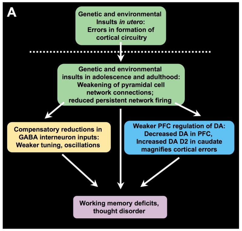
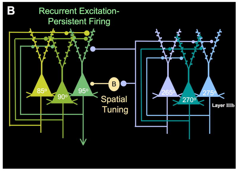
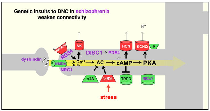
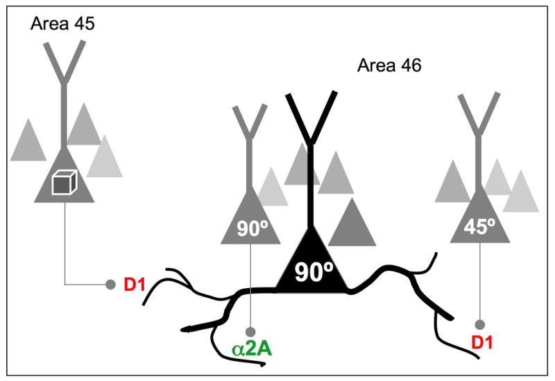
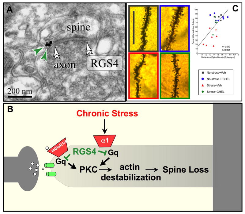
The symptoms of schizophrenia involve profound dysfunction of the prefrontal cortex (PFC). PFC networks create our "mental sketch pad", and PFC dysfunction contributes to symptoms such as cognitive deficits, thought disorder, delusions and hallucinations. Neuropathological studies of schizophrenia have shown marked loss of dendritic spines in deep layer III, the sublayer where PFC microcircuits reside. The microcircuits consist of recurrent excitatory pyramidal cell networks that interconnect on spines, and excite each other via NMDA receptor signaling. The pyramidal cell persistent firing is sculpted by lateral inhibition from GABAergic basket and chandelier cells, thus creating tuned, persistent firing needed for accurate representational knowledge (i.e., working memory). The strength of pyramidal cell network connections is markedly and flexibly altered by intracellular signaling pathways in dendritic spines, a process called dynamic network connectivity (DNC). DNC proteins such as HCN channels are concentrated on dendritic spines in deep layer III. Under optimal conditions, network inputs to pyramidal cells are strengthened by noradrenergic alpha-2A inhibition of cAMP-HCN channel signaling, and sculpted by dopamine D1-cAMP-HCN channel weakening of inappropriate inputs. However, with stress exposure, high levels of cAMP-HCN channel signaling produces a collapse in network firing. With chronic stress exposure, spines reduce in size and are lost, and this process involves increased PKC signaling. Importantly, molecules that normally strengthen PFC networks connections and/or reverse the stress response, are often genetically altered in schizophrenia. As exposure to stress is a key factor in the precipitation of schizophrenic symptoms, these dysregulated signaling pathways in deep layer III may interact with already vulnerable circuitry to cause spine loss and the descent into illness.


Figures

Figure 1
Figure 1
Prefrontal cortical circuitry and schizophrenia. A. An overview of a possible progression of brain changes in schizophrenia. Genetic and/or environmental insults alter the precise formation of cortical pyramidal cell microcircuits in utero. These insults continue to weaken NMDA-mediated, pyramidal cell connections in the maturing brain, particularly during adolescence when there are normal rearrangements of cortical circuitry. Layer III pyramidal cell recurrent microcircuits may be particularly vulnerable, as this layer is the focus of spine loss in schizophrenia. Loss of NMDA microcircuit activity leads to a variety of compensatory changes, including reduction in GABAergic actions, reduced DA inputs to PFC, and increased DA inputs to caudate. Reductions in GABA may contribute to weaker oscillatory activity in brain. Reductions in both GABA and D1 receptor stimulation in PFC would erode information processing in PFC, increasing inappropriate network inputs. Increased DA inputs in caudate magnify cortical errors to contribute to thought disorder, hallucinations and delusions. Adapted from Lewis and Gonzalez-Burgos, 2006, with emphasis on vulnerability of pyramidal cell recurrent connections. B. The layer III PFC microcircuits that subserve spatial working memory in primate dorsolateral PFC. Based on the work of Goldman-Rakic and colleagues (Goldman-Rakic, 1995).
Figure 1
Figure 1
Prefrontal cortical circuitry and schizophrenia. A. An overview of a possible progression of brain changes in schizophrenia. Genetic and/or environmental insults alter the precise formation of cortical pyramidal cell microcircuits in utero. These insults continue to weaken NMDA-mediated, pyramidal cell connections in the maturing brain, particularly during adolescence when there are normal rearrangements of cortical circuitry. Layer III pyramidal cell recurrent microcircuits may be particularly vulnerable, as this layer is the focus of spine loss in schizophrenia. Loss of NMDA microcircuit activity leads to a variety of compensatory changes, including reduction in GABAergic actions, reduced DA inputs to PFC, and increased DA inputs to caudate. Reductions in GABA may contribute to weaker oscillatory activity in brain. Reductions in both GABA and D1 receptor stimulation in PFC would erode information processing in PFC, increasing inappropriate network inputs. Increased DA inputs in caudate magnify cortical errors to contribute to thought disorder, hallucinations and delusions. Adapted from Lewis and Gonzalez-Burgos, 2006, with emphasis on vulnerability of pyramidal cell recurrent connections. B. The layer III PFC microcircuits that subserve spatial working memory in primate dorsolateral PFC. Based on the work of Goldman-Rakic and colleagues (Goldman-Rakic, 1995).
Figure 2
Figure 2
Some of the DNC signaling pathways that regulate PFC network strength, and their relationship to schizophrenia; mechanisms that strengthen connectivity are shown in green; those that weaken connectivity are shown in red. Genetic alterations in patients with schizophrenia frequently involve DNC proteins that normally serve to strengthen pyramidal cell connections, and to inhibit stress signaling pathways (shown in purple). Thus, these genetic insults would weaken PFC network connections and increase vulnerability to stress exposure. Adapted from (Arnsten et al., 2010).
Figure 3
Figure 3
Recordings from PFC neurons indicate that network inputs from neurons with similar vs. dissimilar tuning characteristics are modulated differently. Noradrenergic stimulation of α2A-AR strengthens delay-related firing for a neuron's preferred direction, suggesting that these receptors modulate network inputs from neurons with shared stimulus properties (e.g. as shown in this example, strengthening inputs from other 90° neurons). In contrast, dopamine D1 signaling can weaken network connections from neurons with dissimilar characteristics, (e.g. weakening inputs from a 45° neuron). We have proposed that D1 receptor stimulation may also weaken network inputs from other cortical areas, (e.g. visual feature information arriving from area 45), thus determining the breadth of network inputs. Based on (Arnsten et al., 2009).
Figure 4
Figure 4
RGS4 inhibition of PKC signaling may protect PFC gray matter from the detrimental effects of chronic stress exposure. A. RGS4 is abundant in PFC neurons, and is localized near synapses in dendritic spines in monkey dorsolateral PFC (Paspalas et al., 2009). B. A possible signaling mechanism whereby RGS4 inhibits Gq-PKC signaling in spines. Sustained increases in PKC signaling are known to cause collapse of the actin cytoskeleton and spine loss via phosphorylation of MARCKS, which in turn unanchors actin from the membrane (see text). C. Exposure to chronic stress induces working memory deficits and spine loss from layer III PFC pyramidal cells in rats. Inhibition of PKC signaling during stress exposure protects working memory and dendritic spines, and there is a significant correlation between these measures. Thus, loss of RGS4 in schizophrenia may leave patients more vulnerable to spine loss. Black= control conditions; Red= saline treatment during chronic stress exposure; Blue = PKC inhibition under control conditions, Green = PKC inhibition during chronic stress exposure. Adapted from (Hains et al., 2009).

References

    1. Abi-Dargham A, Mawlawi O, Lombardo I, Gil R, Martinez D, Huang Y, Hwang DR, Keilp J, Kochan L, Van Heertum R, Gorman JM, Laruelle M. Prefrontal dopamine D1 receptors and working memory in schizophrenia. J Neurosci. 2002;22:3708–3719. - PMC - PubMed
    1. Akil M, Pierri JN, Whitehead RE, Edgar CL, Mohila C, Sampson AR, Lewis DA. Lamina-specific alterations in the dopamine innervation of the prefrontal cortex in schizophrenic subjects. Am J Psychiatry. 1999;156:1580–1589. - PubMed
    1. Alexander GE, DeLong MR, Strick PL. Parallel organization of functionally segregated circuits linking basal ganglia and cortex. Ann Rev Neurosci. 1986;9:357–381. - PubMed
    1. Arnsten AFT. Stress signaling pathways that impair prefrontal cortex structure and function. Nature Reviews Neuroscience. 2009;32:267–287. - PMC - PubMed
    1. Arnsten AFT, Goldman-Rakic PS. Stress impairs prefrontal cortex cognitive function in monkeys: role of dopamine. Soc Neurosci Abstr. 1990;16:164.

Publication types

MeSH terms