

Rap1 mediates galanin receptor 2-induced proliferation and survival in squamous cell carcinoma
- PMID: 21345369
- PMCID: PMC3090707
- DOI: 10.1016/j.cellsig.2011.02.002
Rap1 mediates galanin receptor 2-induced proliferation and survival in squamous cell carcinoma
Abstract
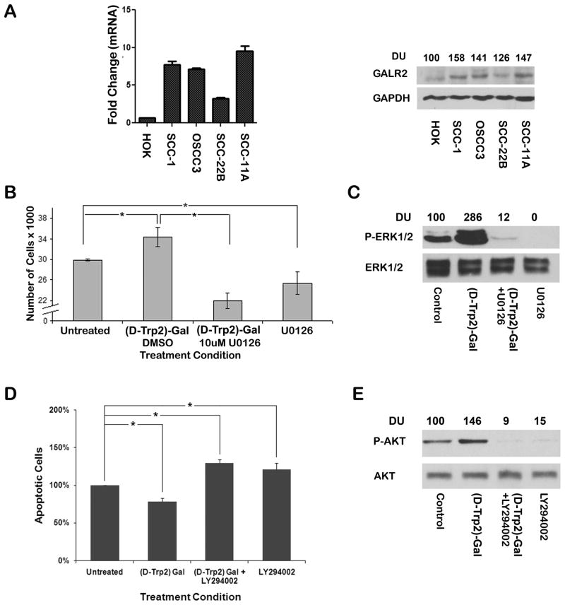
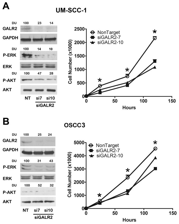
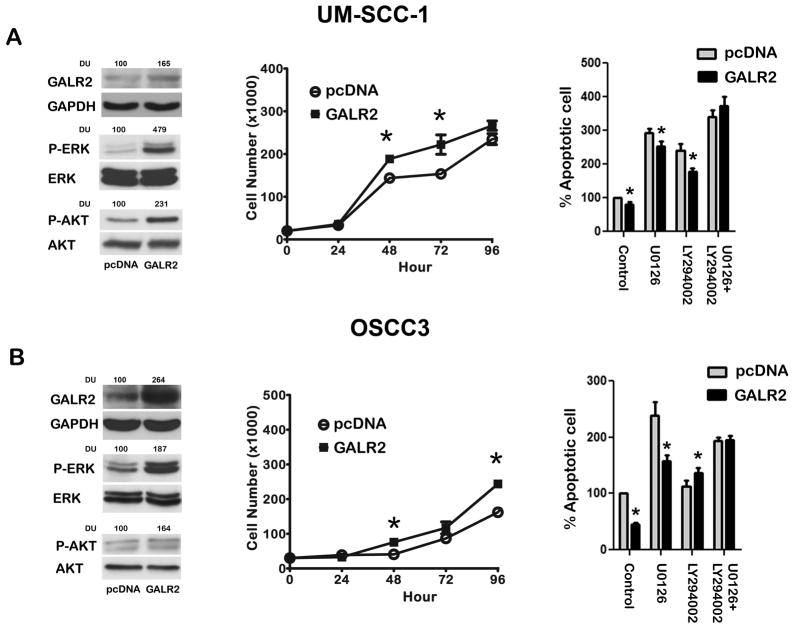
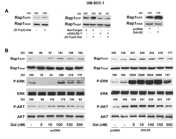
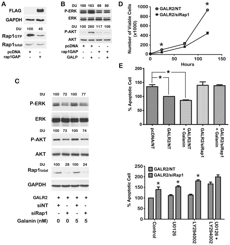
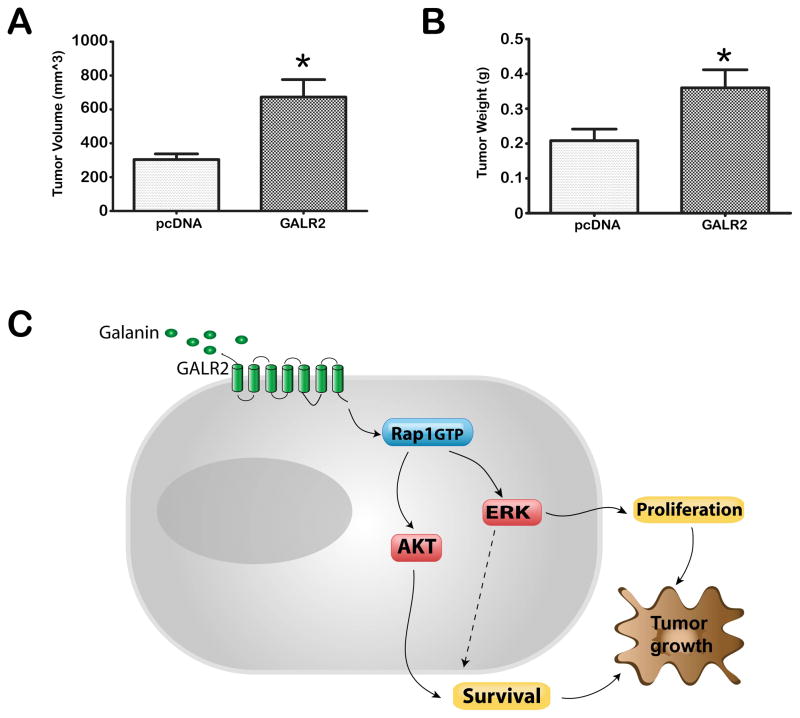
Previously we showed that galanin, a neuropeptide, is secreted by human squamous cell carcinoma of the head and neck (SCCHN) in which it exhibits an autocrine mitogenic effect. We also showed that rap1, a ras-like signaling protein, is a critical mediator of SCCHN progression. Given the emerging importance of the galanin cascade in regulating proliferation and survival, we investigated the effect of GAL on SCCHN progression via induction of galanin receptor 2 (GALR2)-mediated rap1 activation. Studies were performed in multiple SCCHN cell lines by inducing endogenous GALR2, by stably overexpressing GALR2 and by downregulating endogenous GALR2 with siGALR2. Cell proliferation and survival, mediated by the ERK and AKT signaling cascades, respectively, were evaluated by functional and immunoblot analysis. The role of rap1 in GALR2-mediated proliferation and survival was evaluated by modulating expression. Finally, the effect of GALR2 on tumor growth was determined. GALR2 stimulated proliferation and survival via ERK and AKT activation, respectively. Knockdown or inactivation of rap1 inhibited GALR2-induced, AKT and ERK-mediated survival and proliferation. Overexpression of GALR2 promoted tumor growth in vivo. GALR2 promotes proliferation and survival in vitro, and promotes tumor growth in vivo, consistent with an oncogenic role for GALR2 in SCCHN.
Copyright © 2011 Elsevier Inc. All rights reserved.
Conflict of interest statement
Figures
Similar articles
-
Novel anti-tumor mechanism of galanin receptor type 2 in head and neck squamous cell carcinoma cells.Cancer Sci. 2014 Jan;105(1):72-80. doi: 10.1111/cas.12315. Epub 2013 Dec 1. Cancer Sci. 2014. PMID: 24168112 Free PMC article.
-
The G protein-coupled receptor GALR2 promotes angiogenesis in head and neck cancer.Mol Cancer Ther. 2014 May;13(5):1323-33. doi: 10.1158/1535-7163.MCT-13-0904. Epub 2014 Feb 25. Mol Cancer Ther. 2014. PMID: 24568968 Free PMC article.
-
Galanin receptor subtype 2 suppresses cell proliferation and induces apoptosis in p53 mutant head and neck cancer cells.Clin Cancer Res. 2009 Apr 1;15(7):2222-30. doi: 10.1158/1078-0432.CCR-08-2443. Epub 2009 Mar 10. Clin Cancer Res. 2009. PMID: 19276245 Free PMC article.
-
Galanin receptor subtypes 1 and 2 as therapeutic targets in head and neck squamous cell carcinoma.Expert Opin Ther Targets. 2010 Mar;14(3):289-302. doi: 10.1517/14728221003598922. Expert Opin Ther Targets. 2010. PMID: 20148716 Review.
-
Palmitate differentially regulates Spexin, and its receptors Galr2 and Galr3, in GnRH neurons through mechanisms involving PKC, MAPKs, and TLR4.Mol Cell Endocrinol. 2020 Dec 1;518:110991. doi: 10.1016/j.mce.2020.110991. Epub 2020 Aug 22. Mol Cell Endocrinol. 2020. PMID: 32841709 Review.
Cited by
-
Perineural Invasion in Head and Neck Cancer.J Dent Res. 2018 Jul;97(7):742-750. doi: 10.1177/0022034518756297. Epub 2018 Feb 14. J Dent Res. 2018. PMID: 29443582 Free PMC article. Review.
-
Rap1 and its regulatory proteins: the tumor suppressor, oncogene, tumor suppressor gene axis in head and neck cancer.Small GTPases. 2012 Jul-Sep;3(3):192-7. doi: 10.4161/sgtp.20413. Epub 2012 Jun 11. Small GTPases. 2012. PMID: 22684501 Free PMC article.
-
TRIP13 promotes error-prone nonhomologous end joining and induces chemoresistance in head and neck cancer.Nat Commun. 2014 Jul 31;5:4527. doi: 10.1038/ncomms5527. Nat Commun. 2014. PMID: 25078033 Free PMC article.
-
An assembly of galanin-galanin receptor signaling network.J Cell Commun Signal. 2021 Jun;15(2):269-275. doi: 10.1007/s12079-020-00590-3. Epub 2020 Nov 2. J Cell Commun Signal. 2021. PMID: 33136286 Free PMC article.
-
Identification of Gene and MicroRNA Signatures for Oral Cancer Developed from Oral Leukoplakia.Biomed Res Int. 2015;2015:841956. doi: 10.1155/2015/841956. Epub 2015 May 3. Biomed Res Int. 2015. PMID: 26064958 Free PMC article.
References
-
- Kawata M, Matsui Y, Kondo J, Hishida T, Teranishi Y, Takai Y. J Biol Chem. 1988;263(35):18965–18971. - PubMed
-
- Stork PJ. Trends Biochem Sci. 2003;28(5):267–275. - PubMed
-
- D’Silva NJ, Mitra RS, Zhang Z, Kurnit DM, Babcock CR, Polverini PJ, Carey TE. J Cell Physiol. 2003;196(3):532–540. - PubMed
-
- Mitra RS, Zhang Z, Henson BS, Kurnit DM, Carey TE, D’Silva NJ. Oncogene. 2003;22(40):6243–6256. - PubMed
Publication types
MeSH terms
Substances
Grants and funding
LinkOut - more resources
Full Text Sources
Medical
Miscellaneous
