

Quantization and analysis of hippocampal morphometric changes due to dementia of Alzheimer type using metric distances based on large deformation diffeomorphic metric mapping
- PMID: 21345652
- PMCID: PMC3075359
- DOI: 10.1016/j.compmedimag.2011.01.005
Quantization and analysis of hippocampal morphometric changes due to dementia of Alzheimer type using metric distances based on large deformation diffeomorphic metric mapping
Abstract
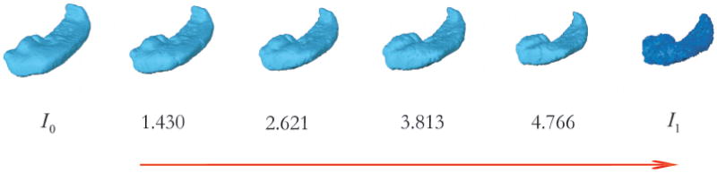
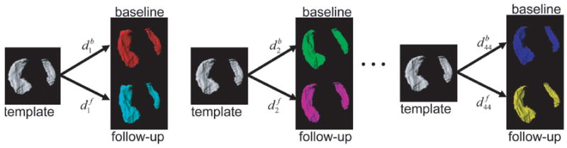
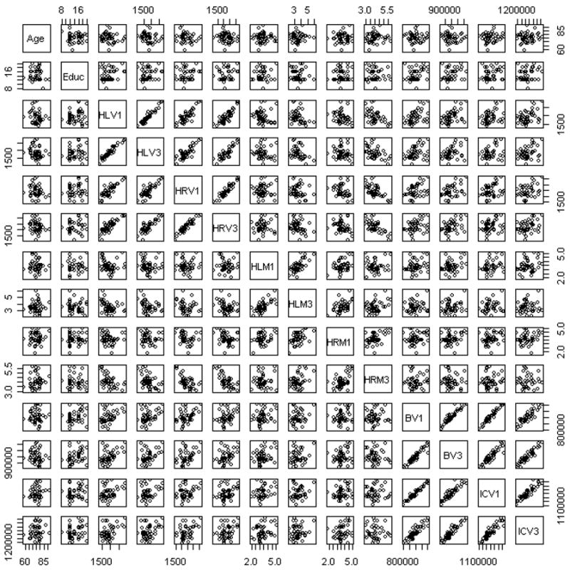
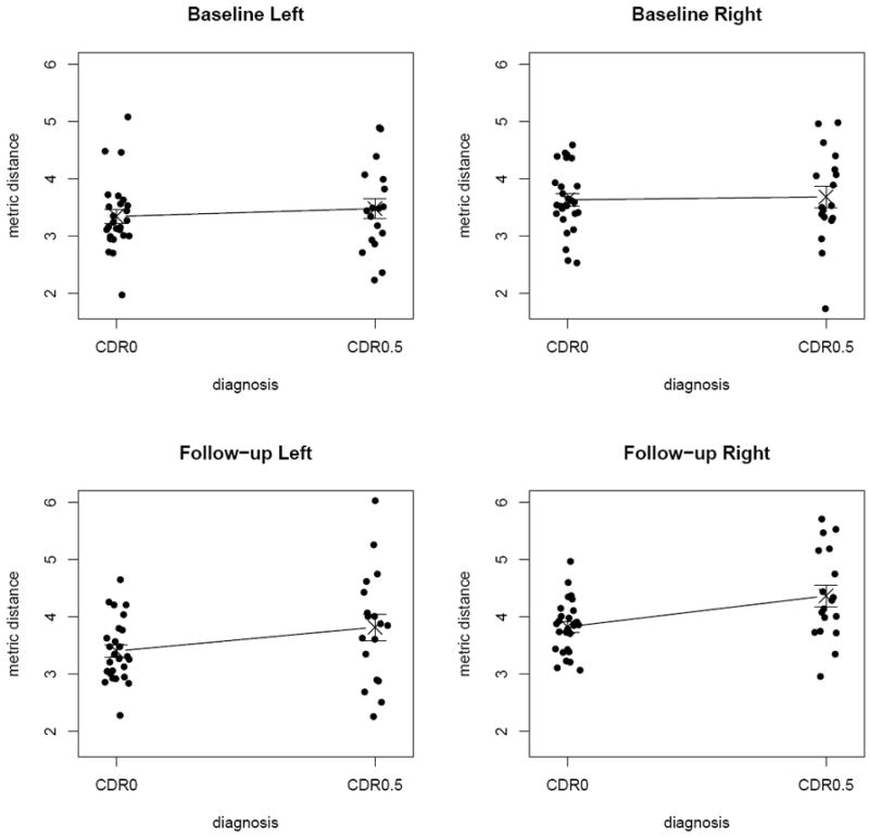
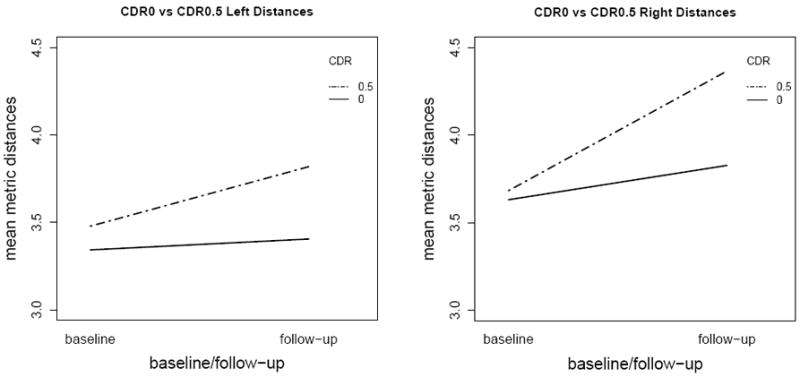
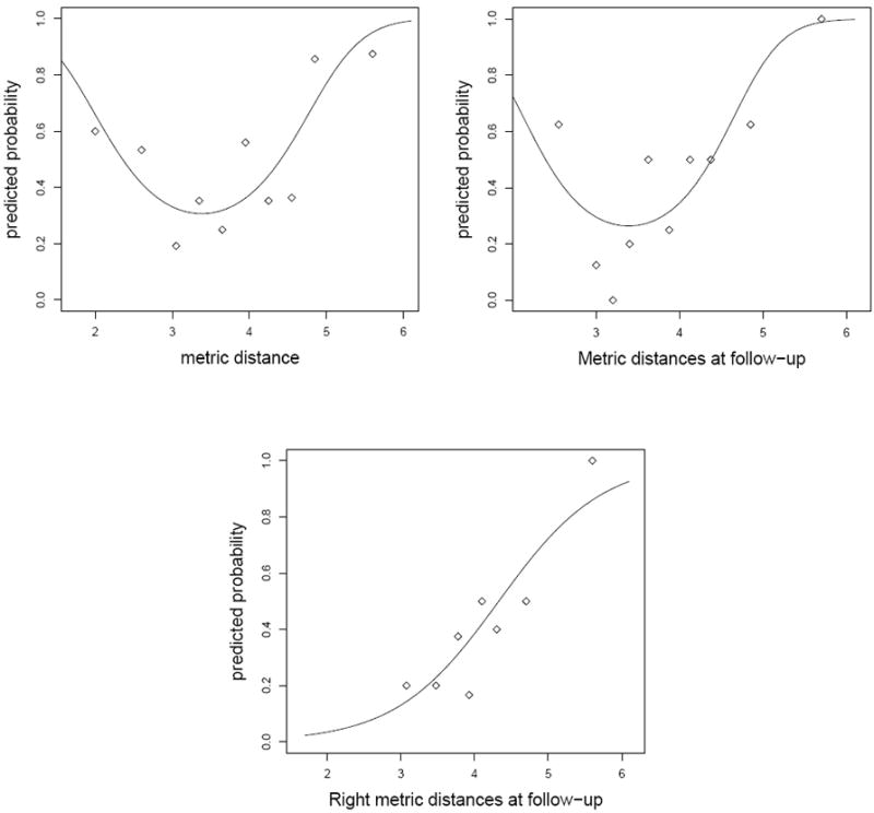
The metric distance obtained from the large deformation diffeomorphic metric mapping (LDDMM) algorithm is used to quantize changes in morphometry of brain structures due to neuropsychiatric diseases. For illustrative purposes we consider changes in hippocampal morphometry (shape and size) due to very mild dementia of the Alzheimer type (DAT). LDDMM, which was previously used to calculate dense one-to-one correspondence vector fields between hippocampal shapes, measures the morphometric differences with respect to a template hippocampus by assigning metric distances on the space of anatomical images thereby allowing for direct comparison of morphometric differences. We characterize what information the metric distances provide in terms of size and shape given the hippocampal, brain and intracranial volumes. We demonstrate that metric distance is a measure of morphometry (i.e., shape and size) but mostly a measure of shape, while volume is mostly a measure of size. Moreover, we show how metric distances can be used in cross-sectional, longitudinal analysis, as well as left-right asymmetry comparisons, and provide how the metric distances can serve as a discriminative tool using logistic regression. Thus, we show that metric distances with respect to a template computed via LDDMM can be a powerful tool in detecting differences in shape.
Copyright © 2011 Elsevier Ltd. All rights reserved.
Figures
References
-
- Hogan RE, Wang L, Bertrand ME, Willmore LJ, Bucholz RD, Nassif AS, Csernansky JG. MRI-based high-dimensional hippocampal mapping in mesial temporal lobe epilepsy. Brain. 2004;127(8):1731–1740. - PubMed
-
- Miller MI. Computational anatomy: shape, growth, and atrophy comparison via diffeomorphisms. Neuroimage. 2004;23(Suppl 1):S19–33. - PubMed
-
- Thompson PM, Hayashi KM, Sowell ER, Gogtay N, Giedd JN, Rapoport JL, de Zubicaray GI, Janke AL, Rose SE, Semple J, Doddrell DM, Wang YL, van Erp TGM, Cannon TD, Toga AW. Mapping cortical change in Alzheimer’s disease, brain development, and schizophrenia. Neuroimage. 2004;23:S2–S18. - PubMed
-
- Grenander U, Miller MI. Computational anatomy: An emerging discipline. Quarterly of Applied Mathematics. 1998;56(4):617–694.
-
- Toga AW. Computational biology for visualization of brain structure. Anatomy and Embryology. 2005;210(5-6):433–438. - PubMed
Publication types
MeSH terms
Grants and funding
LinkOut - more resources
Full Text Sources
Medical
