. 2011 Apr 15;286(15):13430-7.
doi: 10.1074/jbc.M110.205161. Epub 2011 Feb 23.

Structural plasticity of the thioredoxin recognition site of yeast methionine S-sulfoxide reductase Mxr1

Affiliations

Structural plasticity of the thioredoxin recognition site of yeast methionine S-sulfoxide reductase Mxr1

Xiao-Xiao Ma et al. J Biol Chem. .

Abstract

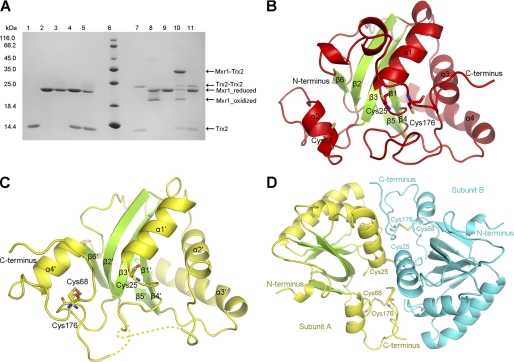
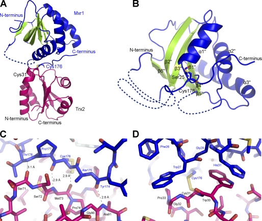
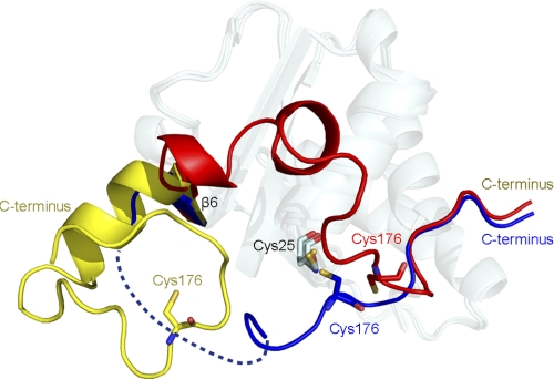
The methionine S-sulfoxide reductase MsrA catalyzes the reduction of methionine sulfoxide, a ubiquitous reaction depending on the thioredoxin system. To investigate interactions between MsrA and thioredoxin (Trx), we determined the crystal structures of yeast MsrA/Mxr1 in their reduced, oxidized, and Trx2-complexed forms, at 2.03, 1.90, and 2.70 Å, respectively. Comparative structure analysis revealed significant conformational changes of the three loops, which form a plastic "cushion" to harbor the electron donor Trx2. The flexible C-terminal loop enabled Mxr1 to access the methionine sulfoxide on various protein substrates. Moreover, the plasticity of the Trx binding site on Mxr1 provides structural insights into the recognition of diverse substrates by a universal catalytic motif of Trx.


Figures

FIGURE 1.
FIGURE 1.
A, electrophoresis of the complexes between Trx2C34S and Mxr1 mutants. The Coomassie-stained gel shows the formation of intermolecular disulfide bonds between Trx2C34S and Mxr1 mutants after incubation under non-reducing conditions (see “Experimental Procedures”). The addition of DTT could reduce the disulfide-linked complex (Mxr1-Trx2 and Trx2-Trx2) or the oxidized Mxr1. Lanes 1–5, Trx2C34S, Mxr1C23C176, Mxr1C23C25, Trx2C34S+Mxr1C23C176, and Trx2C34S+Mxr1C23C25 with 5 mm DTT; lane 6, protein marker; lanes 7–11, samples corresponding to lanes 1–5, respectively, without DTT. B, overall structure of the reduced Mxr1. Cys25, Cys68, and Cys176 are shown as sticks. Helices are colored red, and β-strands are green. C, overall structure of Mxr1 in the unusual oxidized form. Cys25, Cys68, and Cys176 are shown as sticks. Helices are colored yellow, and β-strands are green. D, disulfide-linked Mxr1 dimer in the unusual oxidized form. The two Cys25 residues form an intermolecular disulfide bond, whereas Cys68 and Cys176 form an intramolecular disulfide bond.
FIGURE 2.
FIGURE 2.
A, overall structure of the Mxr1-Trx2 complex (Mxr1, blue and green; Trx2, magenta). Cys176 of Mxr1 and Cys31 of Trx2 are labeled as sticks. Helices are colored blue, and β-strands are colored green. B, overall structure of Mxr1 in the Trx2-complexed form. Ser25 (mutation from Cys25) and Cys176 are shown as sticks. Helices are colored blue, and β-strands are green. C, four main-chain hydrogen bonds. D, hydrophobic interactions between the C-terminal loop of Mxr1 (blue) and Trx2 (magenta).
FIGURE 3.
FIGURE 3.
Conformational changes of the C-terminal loop in the three structures (red, reduced form; yellow, oxidized form; blue, Trx2-complexed form). The core structures are treated with the semitransparent effect.
FIGURE 4.
FIGURE 4.
Superposition of the free (magenta) and Trx-complexed (green) substrate proteins. A, 1Z5Y/1JPE (DsbD from Escherichia coli). B, 2IWT/1AVA (barley alpha-amylase/subtilisin inhibitor from Hordeum vulgare). C, 2K9F/2K0R (DsbD from Neisseria meningitidis). D, 2PUK/2PVD (ferredoxin-thioredoxin reductase from Synechocystis sp.). E, 2O8V/1SUR (3′-Phosphoadenosine-5′-phosphosulfate reductase from Escherichia coli). F, 2IPA/1Z2D (ArsC, arsenate reductase from Bacillus subtilis).

References

    1. Arnér E. S., Holmgren A. (2000) Eur. J. Biochem. 267, 6102–6109 - PubMed
    1. Weissbach H., Etienne F., Hoshi T., Heinemann S. H., Lowther W. T., Matthews B., St. John G., Nathan C., Brot N. (2002) Arch. Biochem. Biophys. 397, 172–178 - PubMed
    1. Moskovitz J., Weissbach H., Brot N. (1996) Proc. Natl. Acad. Sci. U.S.A. 93, 2095–2099 - PMC - PubMed
    1. Moskovitz J., Berlett B. S., Poston J. M., Stadtman E. R. (1997) Proc. Natl. Acad. Sci. U.S.A. 94, 9585–9589 - PMC - PubMed
    1. Kallis G. B., Holmgren A. (1980) J. Biol. Chem. 255, 10261–10265 - PubMed

Publication types

MeSH terms

LinkOut - more resources