. 2011 May;85(10):5125-35.
doi: 10.1128/JVI.01682-10. Epub 2011 Feb 23.

Cytomegalovirus UL103 controls virion and dense body egress

Affiliations

Cytomegalovirus UL103 controls virion and dense body egress

Jenny Ahlqvist et al. J Virol. 2011 May.

Abstract

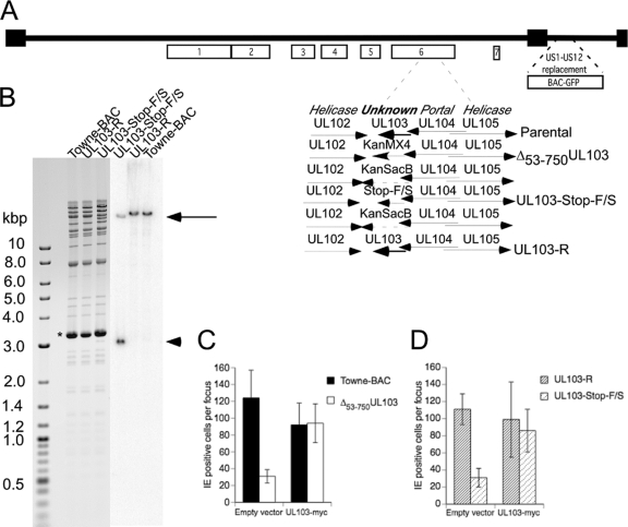
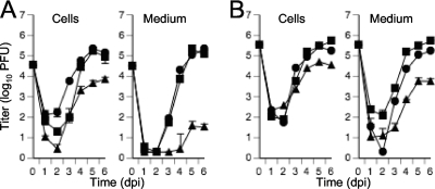
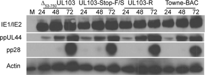
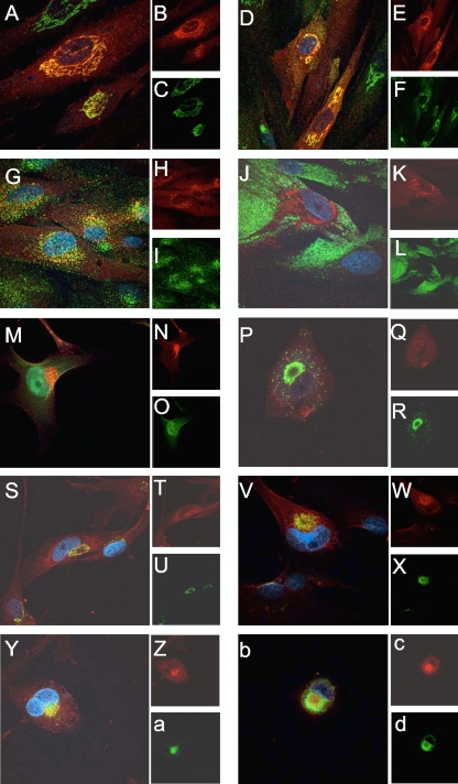
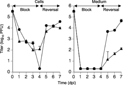
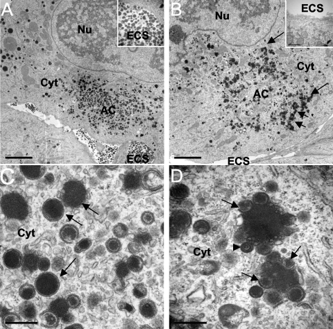
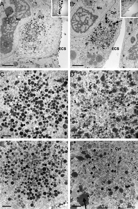
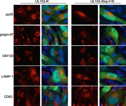
Human cytomegalovirus UL103 encodes a tegument protein that is conserved across herpesvirus subgroups. Mutant viruses lacking this gene product exhibit dramatically reduced accumulation of cell-free virus progeny and poor cell-to-cell spread. Given that viral proteins and viral DNA accumulate with normal kinetics in cells infected with mutant virus, UL103 appears to function during the late phase of replication, playing a critical role in egress of capsidless dense bodies and virions. Few dense bodies were observed in the extracellular space in mutant virus-infected cells in the presence or absence of the DNA encapsidation inhibitor 2-bromo-5,6-dichloro-1-(β-d-ribofuranosyl)benzimidazole. Upon reversal of encapsidation inhibition, UL103 had a striking impact on accumulation of cell-free virus, but not on accumulation of cell-associated virus. Thus, UL103 plays a novel and important role during maturation, regulating virus particle and dense body egress from infected cells.


Figures

Fig. 1.
Fig. 1.
UL103 mutant virus genome structure and complementation. (A) Schematic depiction of the Towne-BAC genome, where boxes 1 to 7 identify herpesvirus-conserved gene clusters (31). The replacement of the US1-US12 region in Towne-BAC with a BAC and a GFP eukaryotic expression cassette has been previously described (30). The region encompassing UL103 in gene cluster 6 is shown expanded, with Towne-BAC, rescue, intermediate Kanr-SacB constructs, and replacement mutant viruses schematically presented. (B) Ethidium bromide-stained electrophoretically separated AvrII-NheI restriction fragment digestion products of Towne-BAC, UL103-R, and UL103-Stop-F/S Bacmid DNA (left panel) and the same samples following DNA blot hybridization to detect the UL103 ORF (right panel). Insertion of AvrII restriction sites into the UL103-Stop-F/S virus resulted in a band at 18.9 kbp (black arrow) and one at 3 kbp (black arrowhead). The thick band at 3.3 kbp (asterisk) is plasmid pSim6. (C and D) Complementation assay using UL103myc-expressing and control HF. Towne-BAC, Δ53-750UL103, UL103-R, and UL103-Stop-F/S virus infections were carried out at an MOI of 0.005 in the presence of 0.01% CMV antibody-positive pooled human IgG to prevent secondary plaque formation. Cells were fixed and stained for IE1/IE2 at 7 days postinfection (dpi), and results are graphed as the average numbers of cells per focus ± standard deviation (SD) (error bars).
Fig. 2.
Fig. 2.
Multistep (A) (MOI of 0.3) or single-step (B) (MOI of 3) growth curves of Towne-BAC (squares), UL103-R (circles), and UL103-Stop-F/S (triangles) reconstituted viruses. The cells and medium were harvested at the indicated times postinfection and frozen at −80°C. The virus titer was determined in triplicate by plaque assay on HF and graphed as log10 mean value ± SD. The data point at 0 dpi indicates the input virus dose.
Fig. 3.
Fig. 3.
Immunoblot analyses of viral proteins from HF infected with Towne-BAC, UL103-R, UL103-Stop-F/S, or Δ53-750UL103 virus and mock-infected HF (MOI of 3). Detection of IE1/IE2, ppUL44, ppUL99 (pp28), and β-actin were carried out at 24, 48, and 72 hpi. Lane M, mock-infected HF.
Fig. 4.
Fig. 4.
Immunofluorescence localization of cellular and viral antigens in uninfected and UL103myc-infected cells. (A to L) Immunofluorescence localization of cellular antigens in UL103myc-expressing uninfected cells using mouse anti-golgin-97 (C), mouse-anti-GM130 (F), mouse anti-EEA-1 (I), and mouse anti-CD63 (L) and donkey anti-mouse conjugated to FITC (green). UL103-myc (B, E, H, and K) was visualized with chicken anti-myc and donkey anti-chicken secondary antibody conjugated to rhodamine (red). Nuclei were stained with Hoechst 33258 (blue). Panels A, D, G, and J display merged images of Hoechst 33258, rhodamine, and FITC. (M to O) Immunofluorescence localization of UL103myc (N) at 3 dpi of Δ53-750UL103 virus-infected cells (MOI of 1) (O) using chicken anti-myc and anti-chicken secondary antibody conjugated to rhodamine (red). (M) Merged image of rhodamine, GFP fluorescence, and Hoechst 33258. (P to d) Immunofluorescence localization of cellular and viral antigens at 3 dpi of Townevar ATCC-infected cells (MOI of 1). pp28 (R), golgin-97 (U), GM130 (X), EEA-1 (a), and CD63 (d) were detected using donkey anti-mouse secondary antibody conjugated to FITC, while myc (Q, T, W, Z, and c) was detected with donkey anti-chicken secondary antibody conjugated to rhodamine. Panels P, S, V, Y, and b display merged images of Hoechst 33258, rhodamine, and FITC. UL103myc was functional in these cells, as shown in Fig. 1C and D. Original magnification obtained by confocal microscopy, ×1,000.
Fig. 5.
Fig. 5.
Single-step growth curve (MOI of 3) of UL103-R (circles) and UL103-Stop-F/S (rectangles) viruses during block and reversal of BDCRB. Cells and medium were harvested individually every day for 7 days and frozen at −80°C. Virus titer was determined and graphed as log10 mean value plus SD. The data point at 0 dpi indicates the input virus dose.
Fig. 6.
Fig. 6.
Characterization of viral components in cells treated with BDCRB and infected with UL103-R virus (A and C) or UL103-Stop-F/S virus (B and D) at 4 dpi by TEM. The nucleus (Nu), cytoplasm (Cyt), extracellular space (ECS), and assembly compartment (AC) are indicated. The arrows in panel B point to cytoplasmic aggregates. (C and D) TEM depicting size bar of 0.5 μm. In panels C and D, the arrows point to dense bodies (DBs) and the arrowhead points to a noninfectious enveloped particle (NIEP). Bars, 5 μm (A and B) and 0.5 μm (C and D).
Fig. 7.
Fig. 7.
TEM of cells infected with UL103-R virus (A, C, and E) or with UL103-Stop-F/S virus (B, D, and F) at 5 dpi. (A to D) Virus-infected cells 1 day after the reversal (5 dpi) of BDCRB. (E and F) Virus-infected cells that were not treated with BDCRB. The small black arrows point to viral particles, and the black arrowheads point to DBs. The large black arrows in panels B and F point to aggregates. The nucleus (Nu), cytoplasm (Cyt), and extracellular space (ECS) are indicated. Bars, 5 μm (A and B) and 1 μm (C to F).
Fig. 8.
Fig. 8.
Immunofluorescence localization of viral and cellular antigens in cells infected with UL103-R or UL103-Stop-F/S virus and treated with BDCRB. The cells were infected at an MOI of 0.3 and fixed 4 dpi. pp28, golgin-97, GM130, LAMP-1, and CD63 were visualized with donkey anti-mouse conjugated to rhodamine. Original magnification, ×1,000.

Similar articles

Cited by

References

    1. Abate D. A., Watanabe S., Mocarski E. S. 2004. Major human cytomegalovirus structural protein pp65 (ppUL83) prevents interferon response factor 3 activation in the interferon response. J. Virol. 78:10995–11006 - PMC - PubMed
    1. Adamo J. E., Schroer J., Shenk T. 2004. Human cytomegalovirus TRS1 protein is required for efficient assembly of DNA-containing capsids. J. Virol. 78:10221–10229 - PMC - PubMed
    1. AuCoin D. P., Smith G. B., Meiering C. D., Mocarski E. S. 2006. Betaherpesvirus-conserved cytomegalovirus tegument protein ppUL32 (pp150) controls cytoplasmic events during virion maturation. J. Virol. 80:8199–8210 - PMC - PubMed
    1. Baldick C. J., Jr., Shenk T. 1996. Proteins associated with purified human cytomegalovirus particles. J. Virol. 70:6097–6105 - PMC - PubMed
    1. Bartz S. R., Vodicka M. A. 1997. Production of high-titer human immunodeficiency virus type 1 pseudotyped with vesicular stomatitis virus glycoprotein. Methods 12:337–342 - PubMed

Publication types

LinkOut - more resources