. 2011 May;85(10):5183-96.
doi: 10.1128/JVI.02302-10. Epub 2011 Feb 23.

TRIM22 inhibits HIV-1 transcription independently of its E3 ubiquitin ligase activity, Tat, and NF-kappaB-responsive long terminal repeat elements

Affiliations

TRIM22 inhibits HIV-1 transcription independently of its E3 ubiquitin ligase activity, Tat, and NF-kappaB-responsive long terminal repeat elements

Anna Kajaste-Rudnitski et al. J Virol. 2011 May.

Erratum in

Abstract

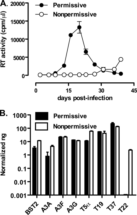
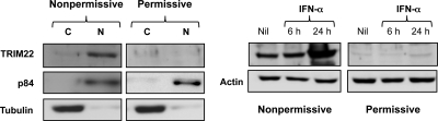
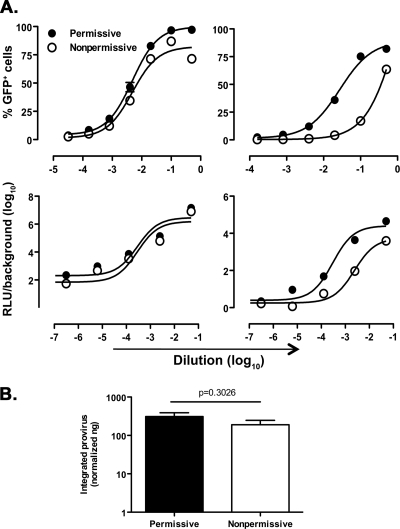
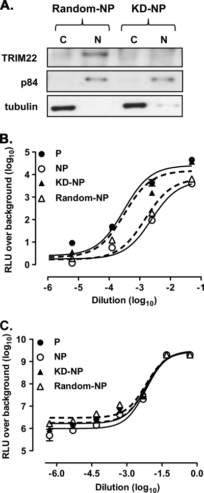
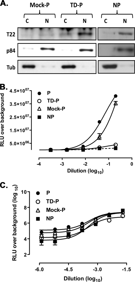
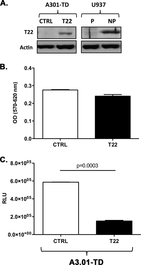
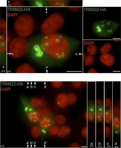
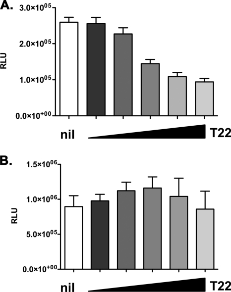
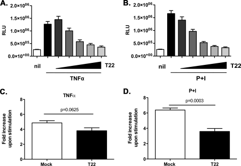
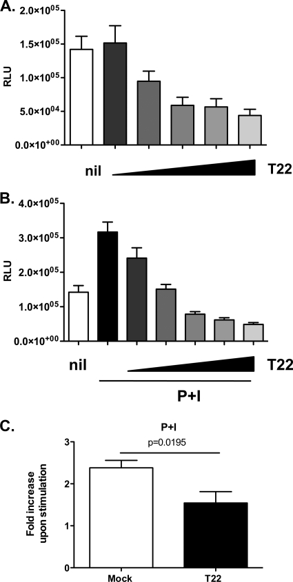
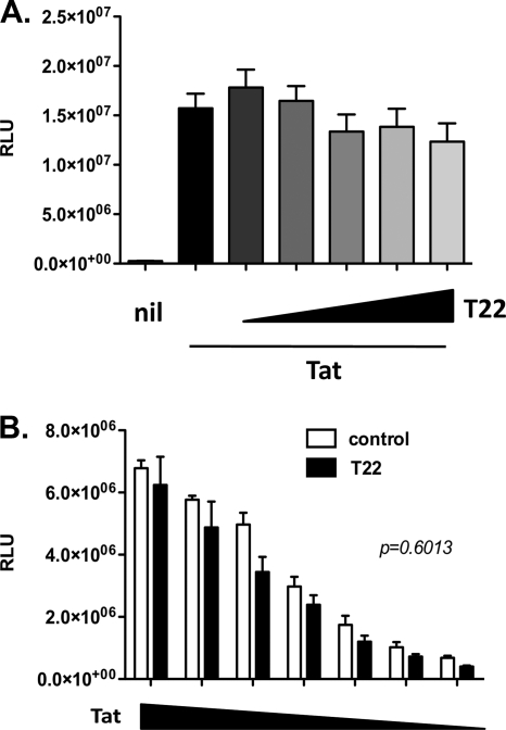
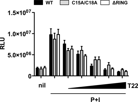
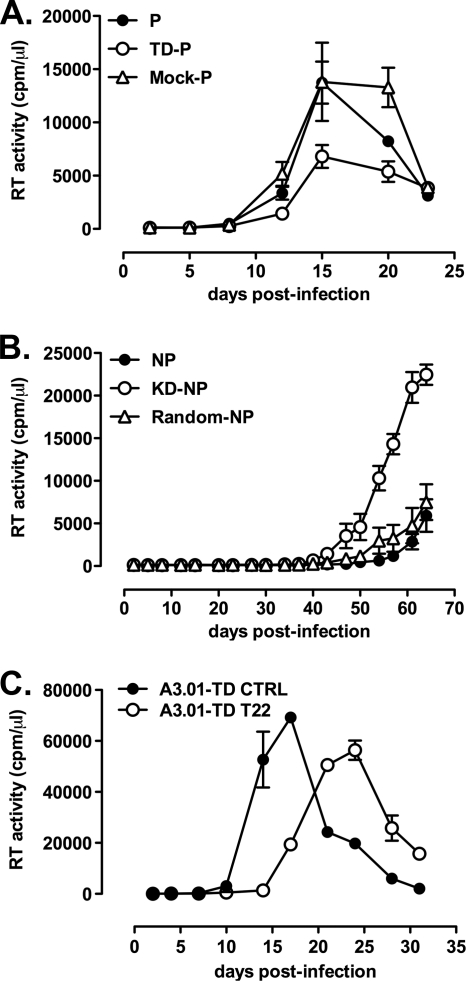
Previous studies identified clones of the U937 promonocytic cell line that were either permissive or nonpermissive for human immunodeficiency virus type 1 (HIV-1) replication. These clones were investigated further in the search for host restriction factors that could explain their differential capacity to support HIV-1 replication. Among known HIV-1 restriction factors screened, tripartite motif-containing protein 22 (TRIM22) was the only factor constitutively expressed in nonpermissive and absent in permissive U937 cells. Stable TRIM22 knockdown (KD) rescued HIV-1 long-terminal-repeat (LTR)-driven transcription in KD-nonpermissive cells to the levels observed in permissive cells. Conversely, transduction-mediated expression of TRIM22 in permissive cells reduced LTR-driven luciferase expression by ∼7-fold, supporting a negative role of TRIM22 in HIV-1 transcription. This finding was further confirmed in the human T cell line A3.01 expressing TRIM22. Moreover, overexpression of TRIM22 in 293T cells significantly impaired basal and phorbol myristate acetate-ionomycin-induced HIV-1 LTR-driven gene expression, whereas inhibition of tumor necrosis factor alpha-induced viral transcription was a consequence of lower basal expression. In agreement, TRIM22 equally inhibited an LTR construct lacking the tandem NF-κB binding sites. In addition, TRIM22 did not affect Tat-mediated LTR transactivation. Finally, these effects were independent of TRIM22 E3 ubiquitin-ligase activity. In the context of replication-competent virus, significantly higher levels of HIV-1 production were observed in KD-nonpermissive versus control nonpermissive U937 cells after infection. In contrast, lower peak levels of HIV-1 replication characterized U937 and A3.01 cells expressing TRIM22 versus their control transduced counterpart. Thus, nuclear TRIM22 significantly impairs HIV-1 replication, likely by interfering with Tat- and NF-κB-independent LTR-driven transcription.


Figures

Fig. 1.
Fig. 1.
Nonpermissive, but not permissive, U937 cell lines characterized by inefficient HIV-1 replication selectively express endogenous TRIM22. (A) Efficient and inefficient X4 HIV-1 replication in U937 cell lines. Permissive (•) and nonpermissive (○) cells were infected with HIV-1IIIB at an MOI of 1. The kinetics of viral replication were determined by measuring the RT activity in the supernatant of infected cultures harvested every 3 to 4 days. The data are expressed as means ± the standard errors of the mean (SEM; n = 3 replicas) of one representative of two independent experiments. (B) Semiquantitative real-time PCR was performed on total cDNA from unstimulated permissive and nonpermissive cell clones, normalized for GAPDH expression. The results are expressed as the normalized ng obtained through extrapolation from relative standard curves for each mRNA established from total cDNA of IFN-α treated nonpermissive cells. The results are expressed as means ± the SEM of one representative of three independent experiments performed in duplicates.
Fig. 2.
Fig. 2.
Nuclear expression and IFN-α induction of TRIM22 in nonpermissive, but not permissive, U937 cell lines. Selective TRIM22 expression was observed in the nucleus (N) but not in the cytoplasm (C) of U937 nonpermissive cells (left panel). Cytoplasmic and nuclear fractions of nonpermissive and permissive U937 protein extracts were subjected to SDS-PAGE and blotted with a polyclonal rabbit serum raised against TRIM22. Immunoblots against cytoplasmic α-tubulin and nuclear p84 antigens served as controls of the efficient cell compartment separation. The WCE obtained from nonpermissive and permissive U937 cells were obtained after 6 and 24 h of stimulation with 1,000 U of IFN-α/ml, evaluated by SDS-PAGE, and blotted with a polyclonal rabbit serum raised against TRIM22 (right panel). Actin was used as a control.
Fig. 3.
Fig. 3.
Selective impairment of HIV-1 LTR-driven gene expression in nonpermissive U937 cells. (A) Permissive and nonpermissive U937 cells were transduced with serially diluted minHIVGFP, HIVGFP, minHIVLuc, or HIVLuc single-round reporter viruses. The percentages of GFP+ cells or RLU were measured 72 h after transduction. Titration curves were obtained by nonlinear regression curve fit using Prism GraphPad software v4.0. The results are representative of three independently performed experiments, expressed as means ± the SEM (n = 3 replicas). The 50% efficacy concentrations expressed in log10 EC50 and their respective P values are reported in Table 2. (B) Similar levels of proviral HIV DNA integration in permissive and nonpermissive U937 cells infected with HIVGFP were observed. Semiquantitative Alu-PCR was performed on genomic DNA extracted from permissive and nonpermissive U937 cells infected with VSV-G-pseudotyped HIVGFP at the dilution 1:10, which yields a major discrepancy in the percentage of GFP+ cells between permissive and nonpermissive cells. Cells were maintained in culture for 4 weeks before testing; mitochondrial DNA was used as a normalizer. The results are expressed as the normalized ng of integrated transfer vector as described in Materials and Methods. The results were obtained from six independent infections for the permissive and five independent infections for nonpermissive cells and are expressed as means ± the SEM. The P value was calculated using an unpaired Student t test.
Fig. 4.
Fig. 4.
Knockdown of TRIM22 in nonpermissive U937 cells rescues HIV-1 LTR-driven transcription. (A) Stable expression of TRIM22-specific shRNA efficiently inhibits TRIM22 expression in nonpermissive U937 cells. Immunoblots with a polyclonal antibody against TRIM22 of nuclear (N) and cytoplasmic (C) extracts obtained from both random-nonpermissive (Random-NP) and KD-nonpermissive (KD-NP) U937 cells. (B) Rescue of HIV-1 LTR gene expression in KD-nonpermissive cells. Permissive and nonpermissive cells, as well as KD-nonpermissive and random-nonpermissive cells (KD-NP and Random-NP, dashed lines) were infected with serial dilutions of HIVLuc. (C) Comparable levels of SFFV LTR-driven gene expression was observed in permissive and nonpermissive cells, as well as in KD-nonpermissive and random-nonpermissive cells (KD-NP and Random-NP, dashed lines), infected with serial dilutions of minHIVLuc. RLU were quantified at 72 h postinfection, and the EC50s were calculated by using a nonlinear regression curve fit of the Prism GraphPad software v4.0. The results were obtained from a single experiment representative of three independently performed and are expressed as means ± the SEM (n = 3 replicas).
Fig. 5.
Fig. 5.
Transduction of TRIM22 in permissive U937 cells selectively represses HIV-1 LTR-driven luciferase expression. (A) TRIM22 (T22) expression in permissive (P) U937 cells was obtained by cell transduction with a TRIM22-expressing lentiviral vector (TD-P), as shown by immunoblotting of cytoplasmic (C) and nuclear (N) extracts, followed by blotting with an anti-TRIM22 polyclonal Ab. Nonpermissive (NP) cells are shown as a control. (B) TRIM22 inhibits HIV-1 transcription in TRIM22-expressing permissive cells. (C) Comparable levels of SFFV LTR-driven gene expression were observed in permissive and nonpermissive cells, as well as in TD-permissive and mock-permissive cells (TD-P and mock-P), infected with serial dilutions of minHIVLuc. The RLU were quantified at 72 h after infection, and EC50s were calculated by using the nonlinear regression curve fit of the Prism GraphPad software v4.0. The results were obtained from a single experiment representative of three independently performed and are expressed as means ± the SEM (n = 3 replicas).
Fig. 6.
Fig. 6.
Transduction of TRIM22 in A3.01 T cells impairs HIV-1 LTR-driven luciferase expression. (A) TRIM22 (T22) expression in A3.01 cells was obtained by cell transduction with a TRIM22-expressing lentiviral vector (A3.01-TD T22), as shown by immunoblotting of WCE, followed by blotting with an anti-TRIM22 polyclonal Ab. Cells transduced with an empty vector (A3.01-TD CTRL), as well as the U937 permissive (P) and nonpermissive (NP) cells, are shown as a control. (B) Cell viability was measured by an MTT assay in transduced A3.01 cells. (C) TRIM22 inhibits HIV-1 transcription in TRIM22-expressing A3.01 cells infected with 16,000 cpm of minHIVLuc/well. The RLU were quantified at 48 h after infection. The results were obtained from a single experiment representative of three independently performed and are expressed as means ± the SEM (n = 3 replicas). The P value was calculated using an unpaired Student t test.
Fig. 7.
Fig. 7.
TRIM22 forms nuclear and cytoplasmic bodies. HA-tagged TRIM22 was visualized by immunofluorescence 24 h after transfection. TRIM22-HA- and DAPI-stained nuclei were pseudocolored green and red, respectively. The main images represent the extended focus of an entire z-stack. To illustrate the localization of TRIM22 within nuclei, y-z and x-z slices, indicated by arrows, are presented next to images. Two representative fields are shown. Size bars, 10 μm.
Fig. 8.
Fig. 8.
TRIM22 selectively inhibits basal HIV-1 transcription. 293T cells were cotransfected with an HIV-1 LTR-Luc (A) or a CMV-driven Luc (B) reporter, together with increasing amounts of TRIM22-expressing vector (T22, wedges), reaching a maximum of 10-fold excess with respect to the reporter plasmid. RLU were quantified 48 h posttransfection. White columns represent basal reporter activity in the absence of TRIM22 (nil). The results are from three independent experiments performed in triplicates each and expressed as means ± the SEM.
Fig. 9.
Fig. 9.
TRIM22 selectively inhibits the induction of HIV-1 transcription. 293T cells were cotransfected with an HIV-1 LTR-Luc reporter, together with increasing amounts of TRIM22-expressing vector (T22, wedges), reaching a maximum of 10-fold excess in respect to the HIV LTR reporter. At 24 h posttransfection, cells were stimulated with 10 ng of TNF-α/ml (A) or 10 nM PMA and 1 μM ionomycin (P+I) (B) for 16 h. The RLU were quantified at 48 h posttransfection. White columns represent basal reporter activity in the absence of TRIM22 (nil), while the first black bar represents LTR activation upon stimulation, in the absence of TRIM22. Fold inductions upon TNF-α (C) or P+I (D) stimulation were compared in the absence (Mock, white bar) or in the presence of the highest amount of TRIM22 (T22, black bar). The results are from three independent experiments performed in triplicates each and are expressed as means ± the SEM. The P values were calculated using the Mann-Whitney test.
Fig. 10.
Fig. 10.
TRIM22 inhibits the activation of HIV-1 LTR independently of the tandem NF-κB site. 293T cells were cotransfected with an HIV-1 ΔNF-κB LTR-Luc reporter plasmid, together with increasing amounts of TRIM22-expressing vector (T22, wedges), reaching a maximum of 10-fold excess in respect to the HIV LTR reporter. At 24 h posttransfection, the cells were either left unstimulated (A) or were stimulated with 10 nM PMA and 1 μM ionomycin (P+I) (B) for 16 h. The RLU were quantified at 48 h posttransfection. White columns represent the basal reporter activity in the absence of TRIM22 (Nil), while the first black bar in panel B represents LTR activation upon stimulation, in the absence of TRIM22. (C) The fold induction upon P+I stimulation was compared in the absence (Mock, white bar) or in the presence of the highest amount of TRIM22 (T22, black bar). The results are from three independent experiments performed in triplicates each and are expressed as means ± the SEM. The P value was calculated using an unpaired Student t test.
Fig. 11.
Fig. 11.
Tat-mediated HIV-1 LTR transactivation is insensitive to TRIM22. (A) 293T cells were cotransfected with an HIV-1 LTR-Luc reporter plasmid and a fixed amount of Tat-expressing vector, together with increasing amounts of TRIM22-expressing vector (T22, wedges), reaching a maximum of 10-fold excess with respect to the HIV LTR. The RLU were quantified at 48 h posttransfection. White columns represent the basal reporter activity in the absence of TRIM22 (Nil), while the first black bar represents LTR activation upon transactivation, in the absence of TRIM22. (B) 293T cells were cotransfected with an HIV-1 LTR-Luc reporter plasmid and a 10-fold excess of control (control, white bars) or TRIM22-expressing (T22, black bars) vector and decreasing quantities of Tat-expressing vector (Tat, wedges). The RLU were quantified at 48 h posttransfection. The results are from three independent experiments performed in triplicates each and expressed as means ± the SEM. The P value was calculated using an unpaired Student t test.
Fig. 12.
Fig. 12.
TRIM22 inhibits P+I-induced HIV-1 LTR-driven gene expression independently of its E3-ubiquitin ligase activity. 293T cells were cotransfected with an HIV-1 LTR-Luc reporter plasmid and increasing amounts of either wild-type TRIM22-expressing vector (black bars, WT), TRIM22 harboring a functionally defective RING-domain (gray bars, C15A/C18A), or TRIM22 with the RING domain deleted (white bars, ΔRING). At 24 h posttransfection, the cells were stimulated with 10 nM PMA and 1 μM ionomycin (P+I) for 16 h, and the RLU were quantified at 48 h posttransfection. The first columns represent basal reporter activity in the absence of TRIM22 (nil), while the second pair of columns represents LTR activation upon stimulation, in the absence of TRIM22. The results are from three independent experiments performed in triplicates each and expressed as means ± the SEM.
Fig. 13.
Fig. 13.
TRIM22 inhibits HIV-1 replication in U937 and A3.01 cells. (A) Expression of TRIM22 decreases the efficiency of X4 HIV-1 replication in permissive cells (TD-P). (B) Suppression of TRIM22 expression promotes accelerated kinetics and increased peak levels of X4 HIV-1 replication in nonpermissive U937 cells (KD-NP). (C) TRIM22 overexpression leads to delayed viral replication in A3.01 cells (A3.01-T22) compared to mock-transduced controls (A3.01-TD CTRL). Infection supernatants were harvested at different times postinfection, and the RT activity was measured. The results were obtained from a single experiment representative of two independently performed and are expressed as means ± the SEM (n = 3 replicas).

Similar articles

Cited by

References

    1. Alfano M., Rizzi C., Poli G., Biswas P. 2007. HIV-1 Infection of promonocytic U937 and U1 cell lines: clonal distribution of innate restriction factors, p. 89–114 In Herbein G. (ed.), Macrophage and HIV infection. Transworld Research Network, Kerala, India
    1. Asjo B., et al. 1987. Susceptibility to infection by the human immunodeficiency virus (HIV) correlates with T4 expression in a parental monocytoid cell line and its subclones. Virology 157:359–365 - PubMed
    1. Barr S. D., Smiley J. R., Bushman F. D. 2008. The interferon response inhibits HIV particle production by induction of TRIM22. PLoS Pathog. 4:e1000007. - PMC - PubMed
    1. Bates D. L., et al. 2008. Crystal structure of NFAT bound to the HIV-1 LTR tandem κB enhancer element. Structure 16:684–694 - PMC - PubMed
    1. Ben-Neriah Y. 2002. Regulatory functions of ubiquitination in the immune system. Nat. Immunol. 3:20–26 - PubMed

Publication types

Substances

LinkOut - more resources