. 2011 May;85(9):4157-66.
doi: 10.1128/JVI.02449-10. Epub 2011 Feb 23.

Transcriptional repression of C4 complement by hepatitis C virus proteins

Affiliations

Transcriptional repression of C4 complement by hepatitis C virus proteins

Arup Banerjee et al. J Virol. 2011 May.

Abstract

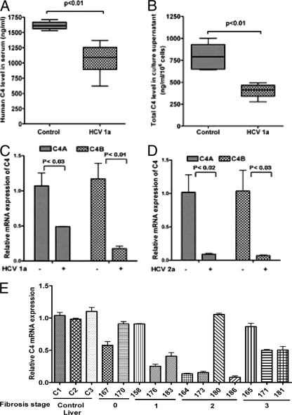
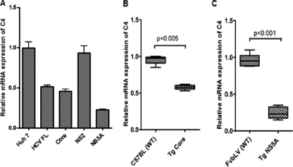
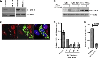
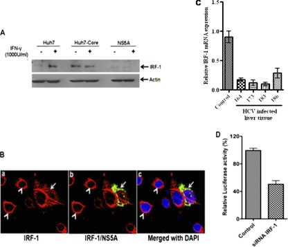
The fourth component of human complement (C4) plays an important role in innate immune function. C4 activity has been observed to be significantly lower in patients with chronic hepatitis C virus (HCV) infections, although the mechanism remains unknown. In this study, we have examined the mechanisms of C4 regulation by HCV. Liver biopsy specimens from patients with chronic HCV infections displayed significantly lower C4 mRNA levels than liver tissue samples from patients with unrelated liver disease. Further, C4 mRNA levels of the two isoforms (C4A and C4B) were significantly reduced in hepatocytes transfected with RNA from HCV genotype 1a or 2a. Subsequently, a significant C4 regulatory role of HCV core or NS5A upon C4 promoter activity was observed. HCV core or NS5A transgenic mice displayed a reduction in C4 mRNA. Gamma interferon (IFN-γ)-induced C4 promoter activation was also impaired in the presence of HCV proteins. We further demonstrated that HCV core reduced the expression of upstream stimulating factor 1 (USF-1), a transcription factor important for basal C4 expression. On the other hand, the expression of interferon regulatory factor 1 (IRF-1), which is important for IFN-γ-induced C4 expression, was inhibited by hepatocytes expressing HCV NS5A. These results underscore the roles of HCV proteins in innate immune regulation in establishing a chronic infection.


Figures

Fig. 1.
Fig. 1.
HCV infection represses C4 expression. (A) There was a significant reduction in total C4 expression displayed in sera from patients infected with HCV genotype 1a (HCV 1a) (n = 12) compared with sera from healthy controls (n = 6). (B) Comparison of total C4 protein level in immortalized human hepatocytes (IHH) and IHH infected with HCV genotype 1a. (C and D) IHH were electroporated with HCV genotype 1a (C) or Huh7.5 cells were electroporated with HCV genotype 2a (D), and the cells were allowed to grow for 6 days. Cellular RNA was extracted, and the levels of mRNA expression of two isoforms of C4 (C4A and C4B) were measured by real-time PCR using specific TaqMan primers and probes. The values are means plus standard deviation (error bars). (E) C4 mRNA expression was measured in HCV genotype 1a-infected paired liver tissue samples (marked by 3-digit numbers) by real-time PCR. Results were normalized to endogenous 18S RNA and compared with non-HCV-infected control liver tissues (C1, C2, and C3). C4 mRNA expression in control liver specimen C1 was arbitrarily set at 1, and the comparative results are shown from individual samples (P < 0.0001 for the values for the group of experimental samples compared to the values for the controls using one-way ANOVA). The values for samples 158, 165, 170, and 180 are not statistically significantly different (P > 0.05) from the values for the controls by Dunnett's multiple-comparison test. The stage of fibrosis of the patient liver is shown at the bottom.
Fig. 2.
Fig. 2.
HCV core and NS5A significantly inhibit C4 promoter activity. (A) HCV polyprotein expression from full-length (FL) genome construct, core, or NS5A significantly suppressed C4 promoter activity in a luciferase reporter assay. However, nonstructural protein NS2 or NS3 did not affect C4 promoter activity. The results obtained were statistically significantly different (P < 0.0001) from the values for the vector control by the unpaired two-tailed t test. (B) Core and NS5A proteins downregulated C4 promoter activity in a dose-dependent manner (0.2 μg and 0.5 μg [the amount is indicated by the height of the black triangle below the bars]). The results obtained were statistically significantly different (P < 0.0001) from the value for the vector control by unpaired two-tailed t test. (C) Core and NS5A proteins exhibited similar levels of suppression of luciferase activity in cells transfected with a plasmid with the wild-type (WT) C4 promoter and in cells transfected with a plasmid in which the C4 promoter had been deleted (pGL3-C4-310). (D) HCV FL, core, or NS5A protein also suppressed IFN-γ-induced C4 promoter activation.
Fig. 3.
Fig. 3.
HCV protein expression downregulates C4 mRNA expression in human hepatocytes and in transgenic mouse liver. (A) Total C4 mRNA level was suppressed in Huh7 cells transfected with HCV FL, core, or NS5A DNA (stable transfectants), while cells transfected with NS2 and used as a negative control did not display any significant effect on C4 mRNA level. GAPDH was used as an endogenous control. C4 mRNA expression in parental hepatocytes (Huh7) was arbitrarily set at 1; the other results shown are from stable transfectants and the values are shown relative to C4 mRNA expression in Huh7 cells (P < 0.0001 for the values for the group of experimental samples using one-way ANOVA). (B and C) C4 mRNA level was measured by real-time PCR and revealed downregulation in transgenic mouse liver expressing HCV core (Tg Core) or NS5A protein (Tg NS5A). Liver RNA from nontransgenic littermates with the same genetic background (C57BL or FvbLV) was used as a control for comparison. Results show the average value of C4 mRNA expression from 6 individual transgenic mice. GAPDH was used an endogenous control.
Fig. 4.
Fig. 4.
USF-1 expression status and C4 promoter activation. (A) Western blot analysis to determine the level of expression of USF-1 in hepatocytes infected with HCV genotype 1a or 2a. (B) Western blot analysis to determine the level of expression of USF-1 in IFN-γ-treated (+) or non-IFN-γ-treated (−) mock-transfected Huh7 cells or Huh7 cells transfected with HCV core or NS5A. Cellular actin was used as an internal control to compare the protein load in each lane. (C) Localization of USF-1 in core-expressing cells by immunofluorescence. Huh7 cells were transiently transfected with HCV core plasmid. The cells were stained after 72 h of transfection with antibody to USF-1 (red) tagged with anti-rabbit Alexa Fluor 594 (a), along with antibody to core (green) tagged with anti-mouse Alexa Fluor 488 (b); merged panels were stained with DAPI (c). Fluorescence, shown in panels a and b, displayed a reduced nuclear expression of USF-1 in HCV core-expressing hepatocytes (white arrows). Cells with no HCV core protein displayed a strong nuclear localization of USF-1 (white arrowheads). (D) USF-1 mRNA expression from selected liver biopsy specimens from HCV-infected patients in comparison to liver biopsy specimens from controls by real-time PCR (P < 0.01 for the values for experimental samples compared to controls using one-way ANOVA). (E) Knockdown of USF-1 by siRNA attenuates IFN-γ-induced C4 promoter activation in Huh7 cells.
Fig. 5.
Fig. 5.
HCV NS5A modulates Stat-1 and suppresses C4 promoter activation. (A) Stat-1 activation was examined by transient transfection of HCV NS5A into Huh7 cells. The inhibitory effect of HCV NS5A on IFN-γ-induced Stat-1 phosphorylation is shown. pStat-1, phosphorylated Stat-1. (B) C4 promoter activity was examined in Stat-1 knockout (KO) (U3A) cells. IFN-γ treatment did not induce C4 promoter activation compared to HT1080 cells. (C) Knockdown of Stat-1 by siRNA attenuates IFN-γ-induced C4 mRNA expression in Huh7 cells.
Fig. 6.
Fig. 6.
IRF-1 expression and C4 promoter activation. (A) Huh7 cells stably transfected with HCV core or NS5A were analyzed for expression of IRF-1 by Western blotting. (B) IRF-1 expression in NS5A-expressing cells by immunofluorescence. Huh7 cells were transiently transfected with HCV NS5A plasmid. (a and b) The cells were stained after 72 h of transfection with antibody to IRF-1 (red) tagged with anti-rabbit Alexa Fluor 594 (a) or antibody to NS5A (green) tagged with anti-mouse Alexa Fluor 488 merged with IRF-1 (b). (c) A reduced expression level of IRF-1 that partially colocalized with NS5A in DAPI-stained cells (shown by arrow) was detected. The absence of HCV NS5A protein was displayed as a clear ring like the perinuclear appearance of IRF-1 (white arrowheads). (C) IRF-1 mRNA expression from selected liver biopsy specimens from HCV-infected patients and comparison to liver biopsy specimens from controls by real-time PCR (P < 0.001 for the values for experimental samples compared to the control values by one-way ANOVA). (D) Knockdown of IRF-1 attenuates IFN-γ-induced C4 promoter activation in Huh7 cells. The results obtained were statistically significantly different (P < 0.0003) from the values for the control by unpaired two-tailed t test.
Fig. 7.
Fig. 7.
Schematic diagram depicting the possible mechanism for HCV core- or NS5A-induced suppression of C4 complement component. HCV core inhibits USF-1 expression, thus inhibiting a basal reduction of C4 expression. On the other hand, IFN-γ-induced C4 activation is attenuated by HCV NS5A via a reduction of IRF-1 expression. P, phosphate.

References

    1. Avirutnan P., et al. 2010. Antagonism of the complement component C4 by flavivirus nonstructural protein NS1. J. Exp. Med. 207:793–806 - PMC - PubMed
    1. Banerjee A., Ray R. B., Ray R. 2010. Oncogenic potential of hepatitis C virus proteins. Viruses 2:2108–2133 - PMC - PubMed
    1. Basu A., Meyer K., Ray R. B., Ray R. 2001. Hepatitis C virus core protein modulates the interferon-induced transacting factors of Jak/Stat signaling pathway but does not affect the activation of downstream IRF-1 or 561 gene. Virology 288:379–390 - PubMed
    1. Basu A., Meyer K., Ray R. B., Ray R. 2002. Hepatitis C virus core protein is necessary for the maintenance of immortalized human hepatocytes. Virology 298:53–62 - PubMed
    1. Blight K. J., Kolykhalov A. A., Rice C. M. 2000. Efficient initiation of HCV RNA replication in cell culture. Science 290:1972–1974 - PubMed

Publication types

LinkOut - more resources