. 2011 May 1;52(6):3368-80.
doi: 10.1167/iovs.10-6991.

Inhibition of Mdm2 sensitizes human retinal pigment epithelial cells to apoptosis

Affiliations

Inhibition of Mdm2 sensitizes human retinal pigment epithelial cells to apoptosis

Sujoy Bhattacharya et al. Invest Ophthalmol Vis Sci. .

Abstract

Purpose: Because recent studies indicate that blocking the interaction between p53 and Mdm2 results in the nongenotoxic activation of p53, the authors sought to investigate whether the inhibition of p53-Mdm2 binding activates p53 and sensitizes human retinal epithelial cells to apoptosis.

Methods: Apoptosis was evaluated by the activation of caspases and DNA fragmentation assays. The Mdm2 antagonist Nutlin-3 was used to dissociate p53 from Mdm2 and, thus, to increase p53 activity. Knockdown of p53 expression was accomplished by using p53 siRNA.

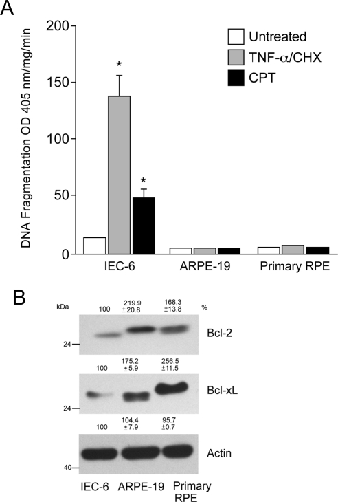
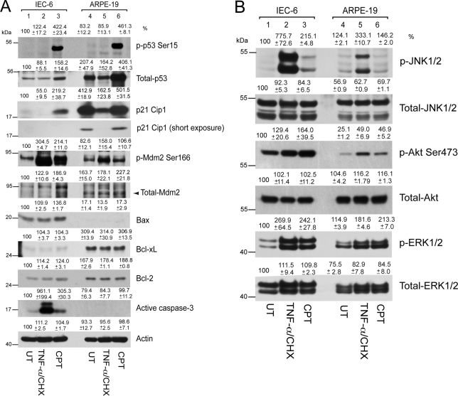
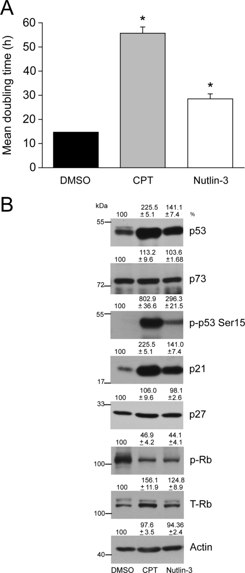
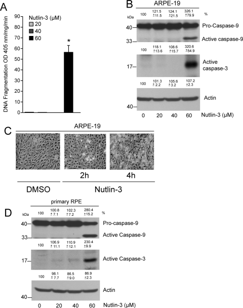
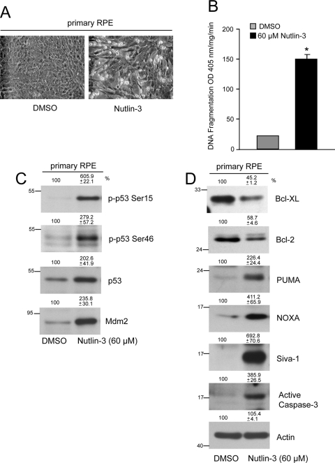
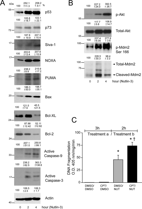
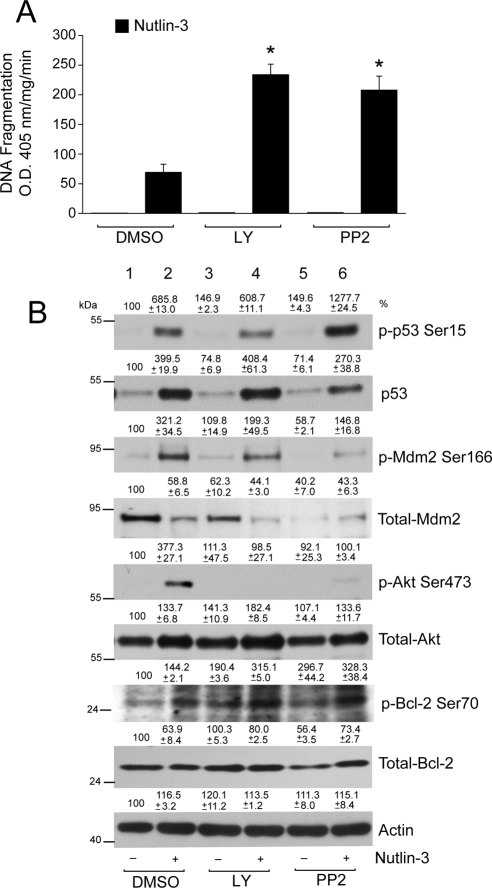
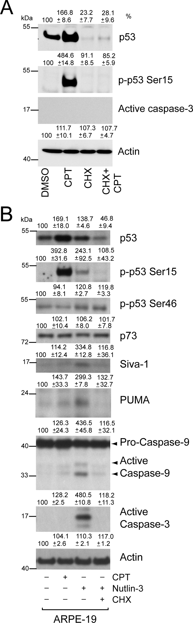
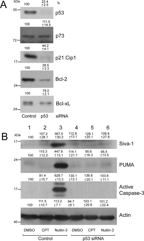
Results: ARPE-19 and primary RPE cells expressed high levels of the antiapoptotic proteins Bcl-2 and Bcl-xL. Exposure of these cells to camptothecin (CPT) or TNF-α/ cycloheximide (CHX) failed to induce apoptosis. In contrast, treatment with the Mdm2 antagonist Nutlin-3 in the absence of CPT or TNF-α/CHX increased apoptosis. Activation of p53 in response to Nutlin-3 also increased levels of Noxa, p53-upregulated modulator of apoptosis (PUMA), and Siva-1, decreased expression of Bcl-2 and Bcl-xL, and simultaneously increased caspases-9 and -3 activities and DNA fragmentation. Knockdown of p53 decreased the basal expression of p21Cip1 and Bcl-2, inhibited the Nutlin-3-induced upregulation of Siva-1 and PUMA expression, and consequently inhibited caspase-3 activation.

Conclusions: These results indicate that the normally available pool of intracellular p53 is predominantly engaged in the regulation of cell cycle checkpoints by p21Cip1 and does not trigger apoptosis in response to DNA-damaging agents. However, the blockage of p53 binding to Mdm2 frees a pool of p53 that is sufficient, even in the absence of DNA-damaging agents, to increase the expression of proapoptotic targets and to override the resistance of RPE cells to apoptosis.


Figures

Figure 1.
Figure 1.
TNF-α/CHX and CPT-mediated apoptosis in IEC-6, ARPE-19, and primary RPE cells. IEC-6, ARPE-19, and primary RPE cells were grown to confluence for 3 days, serum starved for 24 hours, and treated with a combination of TNF-α/CHX or CPT for 4 hours. DNA fragmentation was measured by ELISA (mean ± SEM; n = 3). *P < 0.05; significantly different compared with untreated cells. (B) IEC-6, ARPE-19, and primary RPE cell lysates were analyzed for the levels of Bcl-2 and Bcl-xL using specific antibodies. Actin was used as an internal loading control.
Figure 2.
Figure 2.
TNF-α/CHX- and CPT-mediated signaling in IEC-6 and ARPE-19 cells. IEC-6 and ARPE-19 cells were grown to confluence for 3 days, serum starved for 24 hours, and treated with a combination of TNF-α/CHX or CPT for 4 hours. (A) Cell lysates were separated on SDS-PAGE, and Western blot analysis was carried out using antibodies specific for phospho- and total- p53, Mdm2, p21Cip1, Bax, Bcl-2, Bcl-xL, and active caspase-3. Actin was used as loading control. (B) Cell lysates from this experiment were analyzed for the levels of phospho- and total- JNK1/2, Akt, and ERK1/2 by Western blot analysis.
Figure 3.
Figure 3.
Effect of CPT and Nutlin-3 on the proliferation of ARPE-19 cells. ARPE-19 cells were trypsinized, and equal numbers of cells were seeded in serum-containing medium. Sixteen to 18 hours later, attached cells were treated with 5 μM DMSO, CPT, or Nutlin-3. After treatment, cells were grown for 24 hours and (A) were trypsinized and counted, and the mean doubling time was plotted. *P < 0.05; significantly different compared with DMSO-treated cells. (B) Cell lysates were analyzed by Western blot for the levels of phospho-53 and total p53, Rb, p73, p21Cip1, and p27. Actin was used as an internal loading control.
Figure 4.
Figure 4.
Nutlin-3 induces apoptosis in ARPE-19 and primary RPE cells. (A) ARPE-19 cells were grown to confluence for 3 days, serum starved for 24 hours, and treated with 20, 40, and 60 μM Nutlin-3 for 2 hours. DNA fragmentation was measured by ELISA (mean ± SEM). *P < 0.05; significantly different compared with DMSO. (B) Western blot for active caspase-9 and -3 in 0, 20, 40, and 60 μM Nutlin-3–treated lysates. Actin was used as an internal loading control. (C) ARPE-19 cells were treated with DMSO or Nutlin-3 (60 μM) for 2 and 4 hours, and monolayers were photographed under a phase-contrast microscope equipped with a charge-coupled device camera using Image J software. (D) Primary RPE cultures were grown to confluence, serum starved for 24 hours, and treated with 0, 20, 40, and 60 μM Nutlin-3 for 2 hours. Lysates were analyzed by Western blot for the presence of active caspase-9 and -3. Actin was used as an internal loading control.
Figure 5.
Figure 5.
Activation of p53 signaling in response to Nutlin-3. (A) Primary RPE cells were treated with DMSO or Nutlin-3 (60 μM) for 4 hours, and monolayers were photographed under a phase-contrast microscope equipped with a charge-coupled device camera using ImageJ software. (B) DNA fragmentation was measured by ELISA (mean ± SEM). *P < 0.05; significantly different compared with DMSO. (C) Primary RPE cells were treated with DMSO or 60 μM Nutlin-3, and lysates were analyzed for phospho-p53 (Ser15, Ser46), total p53, and Mdm2. (D) Cell extracts were analyzed to determine the levels of Bcl-xL, Bcl-2, PUMA, Noxa, Siva-1, and active caspase-3 by Western blot analysis. Actin was used as a loading control.
Figure 6.
Figure 6.
Nutlin-3 activates p53 signaling in ARPE-19 cells. (A) ARPE-19 cells were treated with DMSO or Nutlin-3 (60 μM) for 2 and 4 hours. Cell extracts were prepared, and levels of p53, p73, Siva-1, Noxa, PUMA, Bax, Bcl-xL, Bcl-2, active caspase-9, and active caspase-3 proteins were determined using Western blot analysis. Actin was used as an internal loading control. (B) Cell lysates were analyzed for the levels of phospho- and total-Akt and Mdm2 by Western blot analysis. (C) Confluent serum-starved ARPE-19 monolayers were first treated (Treatment a) with DMSO for 3 hours, followed by subsequent treatment (Treatment b) with DMSO (DMSO/DMSO) or (60 μM) Nutlin-3 (DMSO/NUT) for 2 hours. Similarly, cells were treated with (20 μM) CPT for 3 hours, followed by DMSO (CPT/DMSO) or 60 μM Nutlin-3 (CPT/NUT) for 2 hours. DNA fragmentation was measured by ELISA (mean ± SEM; n = 3). *P < 0.05; significantly different compared with DMSO/DMSO. †P < 0.05; significantly different compared with DMSO/Nutlin-3.
Figure 7.
Figure 7.
LY294002 and PP2 sensitizes ARPE-19 cells to Nutlin-3–induced apoptosis. (A) Serum-starved ARPE-19 cell monolayers were pretreated with or without Src kinase inhibitor (PP2; 10 μM) and PI3-kinase inhibitor (LY294002; 10 μM) for 1 hour, followed by treatment with 60 μM Nutlin-3 for 3 hours. An equal volume of DMSO was used as a control. Samples were analyzed for DNA fragmentation using ELISA (mean ± SEM; n = 3). *P < 0.05; significantly different compared with Nutlin-3. (B) Western blot analysis for the levels of phospho- and total- p53, Mdm2, Akt, and Bcl-2 are shown. Actin was used as a loading control.
Figure 8.
Figure 8.
Cycloheximide inhibits Nutlin-3–induced apoptosis. (A) Confluent serum-starved ARPE-19 cells were pretreated with CHX for 1 hour, followed by treatment with or without 20 μM CPT for 3 hours. Western blot for changes in protein levels for phospho-53 and total p53, active caspase-3, and actin are shown. (B) Serum-starved cells were pretreated with CHX for 1 hour, followed by treatment with 60 μM Nutlin-3 for 2 hours. Cells were simultaneously treated with 20 μM CPT for 3 hours, and DMSO-treated cells were used as control. Western blot analysis for changes in protein levels for phospho-53 and total p53, p73, PUMA, Siva-1, and active caspase-9 and caspase-3 are shown.
Figure 9.
Figure 9.
Knockdown of p53 by gene-specific siRNA inhibits Nutlin-3–induced apoptosis. (A) ARPE-19 cells were transfected with control or p53-specific siRNA. Cell lysates were analyzed by Western blot for the levels of p53, p73, p21Cip1, Bcl-2, and Bcl-xL using specific antibodies. (B) Cells transfected with control or p53 siRNA were treated with 20 μM CPT for 3 hours or with 60 μM Nutlin-3 for 2 hours. Cell extracts were analyzed to determine the levels of PUMA, Siva-1, active caspase-3, and actin by Western blot analysis.

References

    1. Strauss O. The retinal pigment epithelium in visual function. Physiol Rev. 2005;85:845–881 - PubMed
    1. Kevany BM, Palczewski K. Phagocytosis of retinal rod and cone photoreceptors. Physiology. 2010;25:8–15 - PMC - PubMed
    1. Pastor JC. Proliferative viteroretinopathy: an overwiew. Surv Ophthalomol. 1998;43:3–18 - PubMed
    1. Lee SC, Known OW, Seong GJ, Kim SH, Ahn JE, Kay ED. Epitheliomesenchymal transdifferentiation of cultured RPE cells. Opthalmic Res. 2001;33:80–86 - PubMed
    1. Ando A, Ueda M, Uyama M, Masu Y, Ito S. Enhancement of dedifferentiation and myoid differentiation of retinal pigment epithelial cells by platelet derived growth factor. Br J Ophthalmol. 2000;84:1306–1311 - PMC - PubMed

Publication types

MeSH terms