. 2011 May;18(5):807-14.
doi: 10.1128/CVI.00434-10. Epub 2011 Feb 23.

Induction of immune tolerance in asthmatic mice by vaccination with DNA encoding an allergen-cytotoxic T lymphocyte-associated antigen 4 combination

Affiliations

Induction of immune tolerance in asthmatic mice by vaccination with DNA encoding an allergen-cytotoxic T lymphocyte-associated antigen 4 combination

Fang Zhang et al. Clin Vaccine Immunol. 2011 May.

Abstract

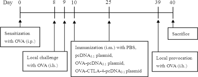
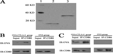
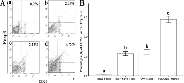
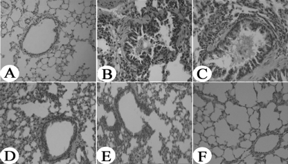
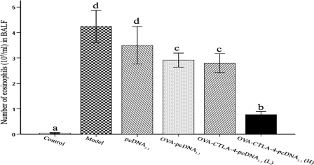
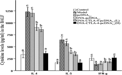
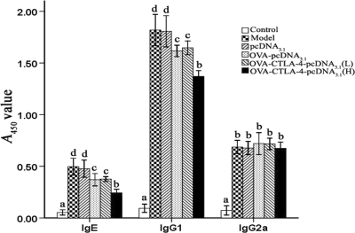
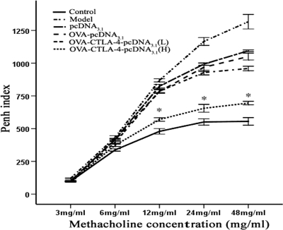
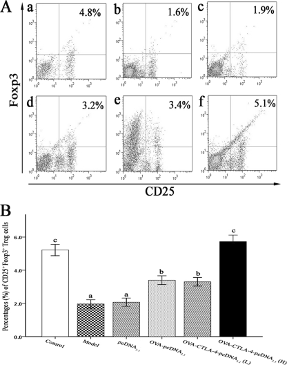
Allergen-specific immunotherapy is a potential treatment for allergic diseases. We constructed an allergen-cytotoxic T lymphocyte-associated antigen 4 (CTLA-4)-encoding DNA vaccine, administered it directly to antigen-presenting cells (APCs), and investigated its ability and mechanisms to ameliorate allergic airway inflammation in an asthmatic mouse model. An allergen-CTLA-4 DNA plasmid (OVA-CTLA-4-pcDNA₃.₁) encoding an ovalbumin (OVA) and the mouse CTLA-4 extracellular domain was constructed and transfected into COS-7 cells to obtain the fusion protein OVA-CTLA-4, which was able to bind the B7 ligand on dendritic cells (DCs), and induced CD25⁺ Foxp3⁺ regulatory T (Treg) cells by the coculture of naive CD4⁺ T cells with DCs in vitro. In an animal study, BALB/c mice were sensitized and challenged with OVA to establish the asthmatic model. Vaccination with a high dose of OVA-CTLA-4-pcDNA₃.₁ significantly decreased interleukin-4 (IL-4) and IL-5 levels and eosinophil counts and prevented OVA-induced reduction of the gamma interferon level in the bronchoalveolar lavage fluid. In addition, these mice suffered less severe airway inflammation and had lower levels of OVA-specific IgE and IgG1 titers in serum. Also, high-dose OVA-CTLA-4-pcDNA₃.₁ vaccination inhibited the development of airway hyperreactivity and prevented OVA-induced reduction of the percentages of Foxp3⁺ Treg cells in the spleen. Our results indicate that a high dose of allergen-CTLA-4-encoding DNA vaccine was more effective in preventing an allergen-induced Th2-skewed immune response through the induction of Treg cells and may be a new alternative therapy for asthma.


Figures

Fig. 1.
Fig. 1.
Immunization protocol.
Fig. 2.
Fig. 2.
OVA-CTLA-4 binds to B7 ligands on DCs in vitro. (A) Purified OVA (lane 2) and OVA-CTLA-4 (lane 3) were determined by Western blot analysis. Lane 1, protein markers. (B) DCs were treated with OVA or OVA-CTLA-4 and lysed for immunoprecipitation with the anti-CD80 antibody. Precipitated proteins were determined by Western blot analysis with anti-OVA antibody. OVA was observed in the OVA-CTLA-4 treated group, whereas no protein was observed in the OVA-treated group. (C) DCs were treated with OVA or OVA-CTLA-4 and lysed for immunoprecipitation with the anti-CD86 antibody. Precipitated proteins were determined by Western blot analysis with anti-OVA antibody. OVA was observed in the OVA-CTLA-4-treated group, while no protein was observed in the OVA-treated group.
Fig. 3.
Fig. 3.
Induction of Treg cells by OVA-CTLA-4 in vitro. (A) Representative results of the percentages of CD25+ Foxp3+ Treg cells from naive CD4+ CD25 T cells only (a) or cocultured with DCs in the presence of medium (b), OVA (c), or OVA-CTLA-4 (d). (B) The percentages of CD25+ Foxp3+ Treg cells were significantly increased in mice from the OVA-CTLA-4-treated group. The values are represent means ± the standard deviations (n = 5). Values with different letters showed significant differences (P < 0.05).
Fig. 4.
Fig. 4.
Reduced lung inflammation in high-dose OVA-CTLA-4-pcDNA3.1-vaccinated mice. Histological findings of lung tissues (original magnification, ×200) in mice from the control group (A), the model group (B), the pcDNA3.1 group (C), the OVA-pcDNA3.1 group (D), the OVA-CTLA-4-pcDNA3.1(L) group (E), and the OVA-CTLA-4-pcDNA3.1(H) group (F) were analyzed by using H&E staining.
Fig. 5.
Fig. 5.
Decreased infiltration of eosinophils into BALF fluid in high-dose OVA-CTLA-4-pcDNA3.1-vaccinated mice. The number of eosinophils in BALF harvested in mice from the control group, the model group, the pcDNA3.1 group, the OVA-pcDNA3.1 group, the OVA-CTLA-4-pcDNA3.1(L) group, and the OVA-CTLA-4-pcDNA3.1(H) group were analyzed by counting. The values represent means ± the standard deviations (n = 5). Values with different letters showed significant differences (P < 0.05).
Fig. 6.
Fig. 6.
Vaccination with a high dose of OVA-CTLA-4-pcDNA3.1 decreased IL-4 and IL-5 levels and prevented the OVA-induced reduction of the IFN-γ level in the BALF. Cytokine production of IL-4, IL-5, and IFN-γ in the BALF from mice in the control group, the model group, the pcDNA3.1 group, the OVA-pcDNA3.1 group, the OVA-CTLA-4-pcDNA3.1(L) group, and the OVA-CTLA-4-pcDNA3.1(H) group was assessed by ELISA. The values represented means ± the standard deviations (n = 5). Values with different letters showed significant differences (P < 0.05).
Fig. 7.
Fig. 7.
Decreased OVA-specific IgE and IgG1 titers in serum in high doses of OVA-CTLA-4-pcDNA3.1-vaccinated mice. The serum levels of OVA-specific IgE, IgG1, and IgG2a in mice from the control group, the model group, the pcDNA3.1 group, the OVA-pcDNA3.1 group, the OVA-CTLA-4-pcDNA3.1(L) group, and the OVA-CTLA-4-pcDNA3.1(H) group were assessed by ELISA. The values represent as means ± the standard deviations (n = 5). Values with different letters showed significant differences (P < 0.05).
Fig. 8.
Fig. 8.
Reduced AHR to cholinergic stimuli in high-doses OVA-CTLA-4-pcDNA3.1-vaccinated mice. Reactivity to methacholine was measured by body plethysmograph, and the data represent the percent increases in Penh values versus baseline values from normal mice at corresponding concentrations of methacholine. Decreased reactivity was seen at all concentrations of methacholine in mice from the OVA-CTLA-4-pcDNA3.1(H) group compared to those from the model group, the pcDNA3.1 group, the OVA-pcDNA3.1 group, and the OVA-CTLA-4-pcDNA3.1(L). *, P < 0.01.
Fig. 9.
Fig. 9.
Vaccination with a high dose of OVA-CTLA-4-pcDNA3.1 prevented the OVA-induced reduction of percentages of CD25+ Foxp3+ Treg cells in the spleen. (A) Representative results of percentages of CD25+ Foxp3+ Treg cells in spleens from mice in the control group (a), the model group (b), the pcDNA3.1 group (c), the OVA-pcDNA3.1 group (d), the OVA-CTLA-4-pcDNA3.1(L) group (e), and the OVA-CTLA-4-pcDNA3.1(H) group (f). (B) The percentages of CD25+ Foxp3+ Treg cells were significantly increased in mice from the OVA-CTLA-4-pcDNA3.1(H) group. The values represented means ± the standard deviations (n = 5). Values with different letters showed significant differences (P < 0.05).

Similar articles

Cited by

References

    1. Abe M., et al. 2008. Foxp3 expression on normal and leukemic CD4+ CD25+ T cells implicated in human T-cell leukemia virus type-1 is inconsistent with Treg cells. Eur. J. Haematol. 81:209–217 - PubMed
    1. Ahrens B., et al. 2009. BCG priming of dendritic cells enhances T regulatory and Th1 function and suppresses allergen-induced Th2 function in vitro and in vivo. Int. Arch. Allergy Immunol. 150:210–220 - PubMed
    1. Akdis M., Akdis C. A. 2007. Mechanisms of allergen-specific immunotherapy. J. Allergy Clin. Immunol. 119:780–791 - PubMed
    1. Cosmi L., et al. 2004. Th2 cells are less susceptible than Th1 cells to the suppressive activity of CD25+ regulatory thymocytes because of their responsiveness to different cytokines. Blood 103:3117–3121 - PubMed
    1. Cusi M. G., et al. 2004. Efficient delivery of DNA to dendritic cells mediated by influenza virosomes. Vaccine 22:735–739 - PubMed

Publication types

MeSH terms