. 2011 Apr;93(4):789-98.
doi: 10.3945/ajcn.110.002766. Epub 2011 Feb 23.

Shmt1 and de novo thymidylate biosynthesis underlie folate-responsive neural tube defects in mice

Affiliations

Shmt1 and de novo thymidylate biosynthesis underlie folate-responsive neural tube defects in mice

Anna E Beaudin et al. Am J Clin Nutr. 2011 Apr.

Abstract

Background: Folic acid supplementation prevents the occurrence and recurrence of neural tube defects (NTDs), but the causal metabolic pathways underlying folic acid-responsive NTDs have not been established. Serine hydroxymethyltransferase (SHMT1) partitions folate-derived one-carbon units to thymidylate biosynthesis at the expense of cellular methylation, and therefore SHMT1-deficient mice are a model to investigate the metabolic origin of folate-associated pathologies.

Objectives: We examined whether genetic disruption of the Shmt1 gene in mice induces NTDs in response to maternal folate and choline deficiency and whether a corresponding disruption in de novo thymidylate biosynthesis underlies NTD pathogenesis.

Design: Shmt1 wild-type, Shmt1(+/-), and Shmt1(-/-) mice fed either folate- and choline-sufficient or folate- and choline-deficient diets were bred, and litters were examined for the presence of NTDs. Biomarkers of impaired folate metabolism were measured in the dams. In addition, the effect of Shmt1 disruption on NTD incidence was investigated in Pax3(Sp) mice, an established folate-responsive NTD mouse model.

Results: Shmt1(+/-) and Shmt1(-/-) embryos exhibited exencephaly in response to maternal folate and choline deficiency. Shmt1 disruption on the Pax3(Sp) background exacerbated NTD frequency and severity. Pax3 disruption impaired de novo thymidylate and purine biosynthesis and altered amounts of SHMT1 and thymidylate synthase protein.

Conclusions: SHMT1 is the only folate-metabolizing enzyme that has been shown to affect neural tube closure in mice by directly inhibiting folate metabolism. These results provide evidence that disruption of Shmt1 expression causes NTDs by impairing thymidylate biosynthesis and shows that changes in the expression of genes that encode folate-dependent enzymes may be key determinates of NTD risk.


Figures

FIGURE 1.
FIGURE 1.
Schematic of folate-mediated one-carbon metabolism in the cytoplasm and nucleus. One-carbon metabolism in the cytoplasm is required for the de novo synthesis of purines and thymidylate and for the remethylation of homocysteine to methionine. One-carbon metabolism in the nucleus synthesizes de novo purine and thymidylate (dTMP) from deoxyuridine monophosphate (dUMP) and serine. THF, tetrahydrofolate; MTHFD1, methylenetetrahydrofolate dehydrogenase; MTR, methionine synthase; MTHFR, methylenetetrahydrofolate reductase; SHMT1, cytoplasmic serine hydroxymethyltransferase; TYMS, thymidylate synthase; DHF, dihydrofolate; DHFR, dihydrofolate reductase; AdoMet, S-adenosylmethionine; AdoHcy, S-adenosylhomocysteine; Sumo, small ubiquitin-like modifier.
FIGURE 2.
FIGURE 2.
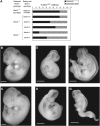
Neural tube defects in Shmt1-deficient embryos. A, B: At gestational day 11.5 (E11.5), +/− embryos from dams fed the diet lacking folate and choline (FCD) exhibited failure of rostral neural tube closure at the midbrain/hindbrain boundary (B). All wild-type littermates (A) were unaffected. Arrows indicate extent of lesions. C, D: At E14.5, affected +/− embryos (D) exhibited prominent exencephaly. Wild-type littermates (C) were unaffected. Arrows indicate extent of lesions. E, F: Hematoxylin and eosin–stained transverse sections of the hindbrain region in E10.5 embryos showed complete failure of closure in the hindbrain region in +/− embryos (F). Wild-type embryos (E) showed normal closure. G, H: Crown-rump length of embryos derived from crosses of Shmt1-deficient mice. Main effects of genotype and diet and the interaction of genotype and diet were determined by mixed-model ANOVA and Tukey's honestly significant difference post hoc test. (G) Crown-rump length as a function of embryonic Shmt1 genotype. n = 90–110 embryos. *Significantly different from +/+, P = 0.057. **Significantly different from +/−, P = 0.007. (H) Crown-rump length as a function of maternal diet. n = 90–120 embryos. **Significantly different from control diet, P ≤ 0.01. The control diet consisted of an experimental feed (AIN93G; Dyets, Bethlehem, PA); FCD refers to an AIN93G diet lacking folate and choline. Scale bars indicate 1 mm.
FIGURE 3.
FIGURE 3.
Metabolic phenotype of the Splotch mutant. A–C: Metabolic phenotype in Splotch mouse embryonic fibroblasts (MEFs). +/+, Sp/+, and Sp/Sp MEF cells were cultured to confluency in α-minimal essential medium supplemented with 14C-deoxyuridine monophosphate (14C-dUMP) and 3H-thymidine or 14C-formate and 3H-hypoxanthine. Efficiency of de novo thymidylate biosynthesis (A) was determined by the relative enrichment of 14C-dUMP (10 μmol/L)/3H-thymidine (500 nmol) in nuclear DNA, and the efficiency of de novo purine biosynthesis (B) was determined by the relative enrichment of 14C-formate (20 μmol/L)/3H-hypoxanthine (2 nmol) in nuclear DNA. All values represent the average of triplicate measures of 3 different cell lines per genotype. (C) The relative ratio of S-adenosylmethionine (AdoMet) to S-adenosylhomocysteine (AdoHcy) was measured in +/+, Sp/+, and Sp/Sp MEF lines. n = 2–3 lines per genotype. Results for all experiments are shown as means ± SDs. All genotype differences were determined by using a Student's t test. *Significantly different from +/+, P ≤ 0.05. **Significantly different from +/+, P < 0.01.
FIGURE 4.
FIGURE 4.
Serine hydroxymethyltransferase (SHMT1) and paired box gene 3 (PAX3) interact in the neural tube. A, B: Protein extracts from gestational day 10.5 (E10.5) embryos isolated from Splotch crosses fed the commercial diet were analyzed by Western blot with a polyclonal anti-SHMT1 antibody (A) and a monoclonal anti-thymidylate synthase (anti-TYMS) antibody (B). Glyceraldehyde 3-phosphate dehydrogenase (GAPDH) was probed with a polyclonal antibody to verify equal loading in both experiments. Relative concentrations of SHMT1 (A) and TYMS (B) were quantified by densitometry with the use of GAPDH as a control. Genotype differences were analyzed by Student's t test with Bonferroni correction (n = 3). Both SHMT1 and TYMS protein concentrations were responsive to the Splotch mutation. *Significantly different from +/+, P < 0.05. **Significantly different from +/+, P ≤ 0.01. C, D: Protein extracts from E8.5 embryos isolated from crosses of +/− dams fed the AIN93G diet lacking folate and choline (FCD diet; Dyets, Bethlehem, PA) were analyzed by Western blot with the use of a polyclonal antibody to PAX3 (C) and TYMS (D). GAPDH and actin were probed to verify equal loading. Relative concentrations of PAX3, SHMT1, and TYMS were quantified by densitometry with the use of GAPDH (C) or actin (D) as a control. Genotype differences for panel C were analyzed by Student's t test with Bonferroni correction (n = 3). Both PAX3 and TYMS protein concentrations were responsive to Shmt1 disruption. *Significantly different from +/+, P < 0.05. E–G: Confocal fluorescent immunohistologic localization of SHMT1 (red) and PAX3 (green) protein in transverse sections at the cephalic level (E), midtrunk (F), and lower trunk (G) in a wild-type E9.0 embryo. Locations of sections are shown in panel H. Scale bars represent 90 μmol/L. nc, neural crest; nt, neural tube; so, somite.
FIGURE 5.
FIGURE 5.
Shmt1 disruption exacerbates neural tube defects in Splotch mutants. A: Incidence of spinal and combined cranial + spinal lesions in Sp/Sp embryos isolated from compound mutant crosses (Pax3,Shmt1) and C57BL/6J-Sp crosses fed a commercial nonpurified rodent diet. B, C: Spina bifida in +/+,+/− (B) and Sp/+,+/− (C) embryos derived from compound mutant crosses fed a commercial nonpurified rodent diet. D, E: Exencephaly in +/+,Shmt1+/− (D) and Sp/+,Shmt1+/− (E) embryos derived from compound mutant crosses fed a commercial nonpurified rodent diet. F, G: Craniorachiscisis in Sp/+,Shmt1−/− (F) and Sp/Sp,Shmt1−/− (G) embryos derived from compound mutant crosses fed the AIN93G diet lacking folate and choline (Dyets, Bethlehem, PA). Scale bars represent 1 mm.

Comment in

References

    1. International Clearinghouse for Birth Defects Monitoring Systems. World atlas of birth defects. 2 ed. Geneva, Switzerland: World Health Organization, 2003
    1. Kirke PN, Molloy AM, Daly LE, Burke H, Weir DG, Scott JM. Maternal plasma folate and vitamin B12 are independent risk factors for neural tube defects. Q J Med 1993;86:703–8 - PubMed
    1. Relton CL, Wilding CS, Laffling AJ, et al. Low erythrocyte folate status and polymorphic variation in folate-related genes are associated with risk of neural tube defect pregnancy. Mol Genet Metab 2004;81:273–81 - PubMed
    1. Christensen B, Arbour L, Tran P, et al. Genetic polymorphisms in methylenetetrahydrofolate reductase and methionine synthase, folate levels in red blood cells, and risk of neural tube defects. Am J Med Genet 1999;84:151–7 - PubMed
    1. Czeizel AE, Dudas I. Prevention of the first occurrence of neural-tube defects by periconceptional vitamin supplementation. N Engl J Med 1992;327:1832–5 - PubMed

Publication types

MeSH terms