

PPARalpha expression protects male mice from high fat-induced nonalcoholic fatty liver
- PMID: 21346097
- PMCID: PMC3056578
- DOI: 10.3945/jn.110.135210
PPARalpha expression protects male mice from high fat-induced nonalcoholic fatty liver
Abstract
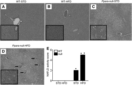
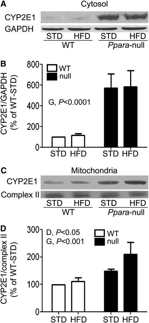
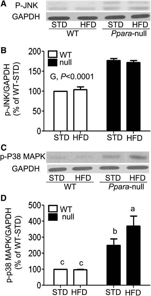
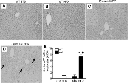
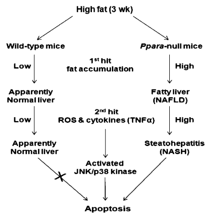
Emerging evidence suggests that the lack of PPARα enhances hepatic steatosis and inflammation in Ppara-null mice when fed a high-fat diet (HFD). Thus, the aim of this study was to determine whether Ppara-null mice are more susceptible to nonalcoholic steatohepatitis (NASH) than their wild-type (WT) counterparts following short-term feeding with a HFD. Age-matched male WT and Ppara-null mice were randomly assigned to consume ad libitum a standard Lieber-DeCarli liquid diet (STD) (35% energy from fat) or a HFD (71% energy from fat) for 3 wk. Liver histology, plasma transaminase levels, and indicators of oxidative/nitrosative stress and inflammatory cytokines were evaluated in all groups. Levels of lobular inflammation and the NASH activity score were greater in HFD-exposed Ppara-null mice than in the other 3 groups. Biochemical analysis revealed elevated levels of ethanol-inducible cytochrome P450 2E1 and TNFα accompanied by increased levels of malondialdehyde as well as oxidized and nitrated proteins in Ppara-null mice. Elevated oxidative stress and inflammation were associated with activation of c-Jun-N-terminal kinase and p38 kinase, resulting in increased hepatocyte apoptosis in Ppara-null mice fed a HFD. These results, with increased steatosis, oxidative stress, and inflammation observed in Ppara-null mice fed a HFD, demonstrate that inhibition of PPARα functions may increase susceptibility to high fat-induced NASH.
Conflict of interest statement
Author disclosures: M. A. Abdelmegeed, S. H. Yoo, L. E. Henderson, F. J. Gonzalez, K. J. Woodcroft, and B. J. Song, no conflicts of interest.
Figures
References
-
- Marchesini G, Bugianesi E, Forlani G, Cerrelli F, Lenzi M, Manini R, Natale S, Vanni E, Villanova N, et al. Nonalcoholic fatty liver, steatohepatitis, and the metabolic syndrome. Hepatology. 2003;37:917–23 - PubMed
-
- Neuschwander-Tetri BA, Caldwell SH. Nonalcoholic steatohepatitis: summary of an AASLD Single Topic Conference. Hepatology. 2003;37:1202–19 - PubMed
-
- Ruhl CE, Everhart JE. Epidemiology of nonalcoholic fatty liver. Clin Liver Dis. 2004;8:501–19 - PubMed
-
- Adams LA, Lymp JF, St Sauver J, Sanderson SO, Lindor KD, Feldstein A, Angulo P. The natural history of nonalcoholic fatty liver disease: a population-based cohort study. Gastroenterology. 2005;129:113–21 - PubMed
-
- Desvergne B, Wahli W. Peroxisome proliferator-activated receptors: nuclear control of metabolism. Endocr Rev. 1999;20:649–88 - PubMed
Publication types
MeSH terms
Substances
Grants and funding
LinkOut - more resources
Full Text Sources
Other Literature Sources
Medical
Molecular Biology Databases
Research Materials
Miscellaneous
