

doi: 10.1126/scitranslmed.3001847.
Tuberculosis immunopathology: the neglected role of extracellular matrix destruction
Affiliations
- PMID: 21346167
- PMCID: PMC3717269
- DOI: 10.1126/scitranslmed.3001847
Item in Clipboard
Tuberculosis immunopathology: the neglected role of extracellular matrix destruction
Sci Transl Med.
.
Abstract
The extracellular matrix in the lung must be destroyed for Mycobacterium tuberculosis--the agent that causes tuberculosis (TB)--to spread. The current paradigm proposes that this destruction occurs as a result of the action of proinflammatory cytokines, chemokines, immune cells, and lipids that mediate TB-associated necrosis in the lung. However, this view neglects the fact that lung matrix can only be degraded by proteases. We propose an original conceptual framework of TB immunopathology that may lead directly to treatments that involve inhibition of matrix metalloproteinase activity to hinder matrix destruction and reduce the morbidity and mortality associated with TB.
Figures
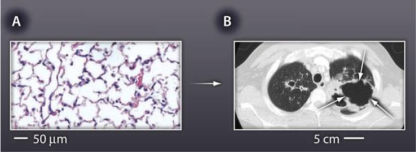
(A) Normal lung architecture is highly organized, supported by an intricate network of extracellular matrix. About 170 alveoli are present per cubic millimeter of healthy lung tissue in humans. (B) This matrix is destroyed during TB, resulting in formation of cavities that are often several centimeters across (white arrows on computerized tomography scan).
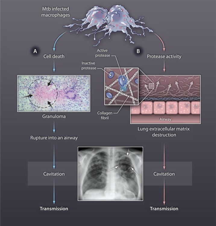
The longstanding paradigm of TB immunopathology (blue arrows) states that cell death leads to the accumulation caseous necrosis in TB granulomas (black arrows), which then rupture into an airway, causing an air-filled cavity in the lung (white arrows on chest radiograph). This model does not explain how the extracellular matrix is degraded. We propose that two independent processes must be taking place: (A) cell death causing the accumulation of caseous material and (B) induction of protease activity to drive destruction of the extracellular matrix, which then results in cavity formation and transmission (red arrows).
References
Publication types
MeSH terms
Grants and funding
LinkOut - more resources
Full Text Sources
Other Literature Sources
Medical
