. 2011 Mar 24;471(7339):508-12.
doi: 10.1038/nature09867. Epub 2011 Feb 23.

Fat cells reactivate quiescent neuroblasts via TOR and glial insulin relays in Drosophila

Affiliations

Fat cells reactivate quiescent neuroblasts via TOR and glial insulin relays in Drosophila

Rita Sousa-Nunes et al. Nature. .

Abstract

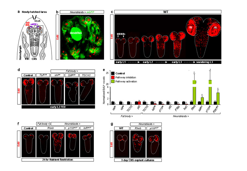
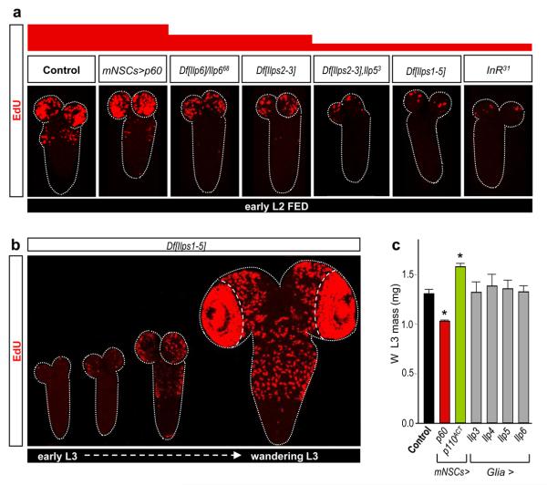
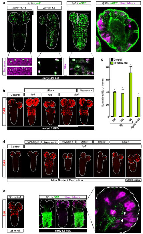
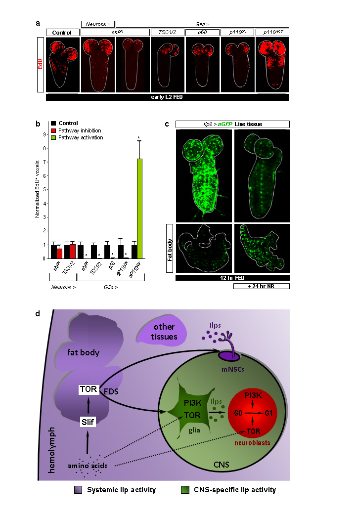
Many stem, progenitor and cancer cells undergo periods of mitotic quiescence from which they can be reactivated. The signals triggering entry into and exit from this reversible dormant state are not well understood. In the developing Drosophila central nervous system, multipotent self-renewing progenitors called neuroblasts undergo quiescence in a stereotypical spatiotemporal pattern. Entry into quiescence is regulated by Hox proteins and an internal neuroblast timer. Exit from quiescence (reactivation) is subject to a nutritional checkpoint requiring dietary amino acids. Organ co-cultures also implicate an unidentified signal from an adipose/hepatic-like tissue called the fat body. Here we provide in vivo evidence that Slimfast amino-acid sensing and Target of rapamycin (TOR) signalling activate a fat-body-derived signal (FDS) required for neuroblast reactivation. Downstream of this signal, Insulin-like receptor signalling and the Phosphatidylinositol 3-kinase (PI3K)/TOR network are required in neuroblasts for exit from quiescence. We demonstrate that nutritionally regulated glial cells provide the source of Insulin-like peptides (ILPs) relevant for timely neuroblast reactivation but not for overall larval growth. Conversely, ILPs secreted into the haemolymph by median neurosecretory cells systemically control organismal size but do not reactivate neuroblasts. Drosophila thus contains two segregated ILP pools, one regulating proliferation within the central nervous system and the other controlling tissue growth systemically. Our findings support a model in which amino acids trigger the cell cycle re-entry of neural progenitors via a fat-body-glia-neuroblasts relay. This mechanism indicates that dietary nutrients and remote organs, as well as local niches, are key regulators of transitions in stem-cell behaviour.


Figures

Fig. 1
Fig. 1. TOR/PI3K signalling in fat body and neuroblasts regulates reactivation
a, Diagram depicting larval fat body (FB) and CNS with central brain (CB), thoracic (Th) and abdominal (Ab) neuromeres, mNSCs, mushroom body (MB NBs) and other neuroblasts (circles) indicated. b, Brain lobe (inset in Fig.1a), showing EdU incorporation in postembryonic neuroblasts (large cells; e.g dotted circle) and their progeny (smaller cells), labelled with nab-GAL4 driving membrane GFP (Neuroblasts>mGFP). c, EdU incorporation timecourse from first-instar (L1) to third-instar (L3) larval stages in the wild-type (WT) CNS (OL, optic lobes). d,f,g, EdU-labelled CNSs from larvae expressing TOR/PI3K components driven by Cg-GAL4 (Fat body>) or nab-GAL4 (Neuroblasts>). e, Histograms of EdU+ voxels from thoracic CNSs of fed larvae, normalized to controls. In this and all subsequent figures, error bars are s.e.m.; * p<0.05. See text, Methods and Supplementary Fig. 2 for details of molecules expressed.
Fig. 2
Fig. 2. Insulin-like peptides but not mNSCs control neuroblast reactivation
a, EdU-labelled CNSs from various Ilp or InR mutants show decreased reactivation whereas larvae with Ilp2-GAL4 driving UAS-p60 (mNSC>p60) do not. b, EdU incorporation timecourse in the CNS of Df[Ilps1-5] larvae. c, The mass of fed L3 larvae at the wandering (W) stage is significantly altered by Ilp2-GAL4 (mNSC>) driving PI3K signalling components but not by repo-GAL4 (Glia>) driving Ilps.
Fig. 3
Fig. 3. CNS-specific Ilps are sufficient for neuroblast reactivation
a, Panels show expression of Ilp3-nLacZ in subsets of neurons (XD311-11) and glia (XD311-1) and Ilp6-GAL4 (Ilp6>nGFP and Ilp6>mGFP) in glia, including BBB surface and cortex glia. b,d EdU-labelled CNSs from larvae overexpressing Ilps in various cell types (see Methods for GAL4 drivers used). c, Histograms of normalized EdU+ voxels in the thoracic CNS for the genotypes in b. e, Ilp6 overexpression in the Ultrabithorax domain (Ubx>Ilp6) reactivates neuroblasts in the normal spatial pattern during NR (left panel). Quiescent/enlarging neuroblasts in the central brain, far from the posterior Ubx domain (middle panels), extend cytoplasmic processes (arrowheads) towards the neuropil, close to long Ubx+ cell processes (right panel). The range of Ilp6 activity is difficult to determine from this experiment. Neurons, glia and neuroblasts are marked by Elav, Repo and Miranda respectively.
Fig. 4
Fig. 4. Ilp6-expressing glia are nutritionally regulated
a, EdU-labelled CNSs from larvae expressing the components indicated. b, Histograms of normalized EdU+ voxels in the thoracic CNS for genotypes in a. c, Ilp6>nGFP expression in the CNS and fat body of fed versus NR larvae. d, Relay model for amino-acid dependent fat body regulation of CNS and body growth. CNS-restricted (green) and systemic (purple) pools of Insulin-like peptides (Ilps) are functionally segregated. Direct amino-acid sensing by glia and neuroblasts may contribute to neuroblast reactivation (dashed arrows). See text for details.

Comment in

References

METHODS REFERENCES

    1. Oldham S, et al. Genetic and biochemical characterization of dTOR, the Drosophila homolog of the target of rapamycin. Genes Dev. 2000;14(21):2689. - PMC - PubMed
    1. Brogiolo W, et al. An evolutionarily conserved function of the Drosophila insulin receptor and insulin-like peptides in growth control. Curr Biol. 2001;11(4):213. - PubMed
    1. Zhang H, et al. Deletion of Drosophila insulin-like peptides causes growth defects and metabolic abnormalities. Proc Natl Acad Sci U S A. 2009;106(46):19617. - PMC - PubMed
    1. Gronke S, et al. Molecular evolution and functional characterization of Drosophila insulin-like peptides. PLoS Genet. 2010;6(2):e1000857. - PMC - PubMed
    1. Colombani J, et al. A nutrient sensor mechanism controls Drosophila growth. Cell. 2003;114(6):739. - PubMed

References

    1. Dhawan J, Rando TA. Stem cells in postnatal myogenesis: molecular mechanisms of satellite cell quiescence, activation and replenishment. Trends Cell Biol. 2005;15:666–73. - PubMed
    1. Coller HA. What's taking so long? S-phase entry from quiescence versus proliferation. Nat Rev Mol Cell Biol. 2007;8:667–70. - PubMed
    1. Yanagida M. Cellular quiescence: are controlling genes conserved? Trends Cell Biol. 2009;19:705–715. - PubMed
    1. Chen E, Finkel T. Preview. The Tortoise, the hare, and the FoxO. Cell Stem Cell. 2009;5:451–2. - PubMed
    1. Sanchez-Garcia I, Vicente-Duenas C, Cobaleda C. The theoretical basis of cancer-stem-cell-based therapeutics of cancer: can it be put into practice? Bioessays. 2007;29:1269–80. - PubMed

Publication types

MeSH terms