. 2011 Jun;131(6):1339-46.
doi: 10.1038/jid.2011.31. Epub 2011 Feb 24.

C/EBPα expression is downregulated in human nonmelanoma skin cancers and inactivation of C/EBPα confers susceptibility to UVB-induced skin squamous cell carcinomas

Affiliations

C/EBPα expression is downregulated in human nonmelanoma skin cancers and inactivation of C/EBPα confers susceptibility to UVB-induced skin squamous cell carcinomas

Elizabeth A Thompson et al. J Invest Dermatol. 2011 Jun.

Abstract

Human epidermis is routinely subjected to DNA damage induced by UVB solar radiation. Cell culture studies have revealed an unexpected role for C/EBPα (CCAAT/enhancer-binding protein-α) in the DNA damage response network, where C/EBPα is induced following UVB DNA damage, regulates the G(1) checkpoint, and diminished or ablated expression of C/EBPα results in G(1) checkpoint failure. In the current study we observed that C/EBPα is induced in normal human epidermal keratinocytes and in the epidermis of human subjects exposed to UVB radiation. The analysis of human skin precancerous and cancerous lesions (47 cases) for C/EBPα expression was conducted. Actinic keratoses, a precancerous benign skin growth and precursor to squamous cell carcinoma (SCC), expressed levels of C/EBPα similar to normal epidermis. Strikingly, all invasive SCCs no longer expressed detectable levels of C/EBPα. To determine the significance of C/EBPα in UVB-induced skin cancer, SKH-1 mice lacking epidermal C/EBPα (CKOα) were exposed to UVB. CKOα mice were highly susceptible to UVB-induced SCCs and exhibited accelerated tumor progression. CKOα mice displayed keratinocyte cell cycle checkpoint failure in vivo in response to UVB that was characterized by abnormal entry of keratinocytes into S phase. Our results demonstrate that C/EBPα is silenced in human SCC and loss of C/EBPα confers susceptibility to UVB-induced skin SCCs involving defective cell cycle arrest in response to UVB.


Conflict of interest statement

CONFLICT OF INTEREST

The authors state no conflict of interest.

Figures

Figure 1
Figure 1. C/EBPα (CCAAT/enhancer-binding protein-α) expression in human skin and precancerous and cancerous lesions
(a) Immunohistochemical (IHC) staining for C/EBPα expression was conducted on 47 individual cases. (b) Expression was scored according to the following: (+ + +) intense nuclear staining in >30% of keratinocytes, (+ +) moderate nuclear staining in 10–30% of keratinocytes, (+) weak nuclear staining in <10% of keratinocytes, and (−) no detectable nuclear staining. Scale bar=20 μm.
Figure 2
Figure 2. C/EBPα (CCAAT/enhancer-binding protein-α) is induced by UVB in human skin in vivo
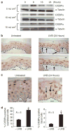
(a) Immunoblot analysis of cell lysates from primary human epidermal keratinocytes. (b) Immunohistochemical (IHC) staining for C/EBPα in sections from untreated and UVB-treated human skin. Human skin biopsies were collected 24 hours after UVB treatment (1 minimum erythemic dose (MED)). (c) C/EBPα-positive staining basal cells in UVB-treated human skin. (d) Percentage of C/EBPα IHC-positive keratinocytes in untreated and UVB-treated human skin. An average of 360 and 400 basal cells and suprabasal cells, respectively, were counted per biopsy. Data are expressed as the mean±SE (N=5 individuals per treatment group). *Significantly different from untreated controls, P<0.025, Student’s t-test. Black arrows indicate C/EBPα-positive basal keratinocytes; dashed line indicates basement membrane. Scale bar=10 μm.
Figure 3
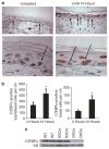
Figure 3. C/EBPα (CCAAT/enhancer-binding protein-α) is induced by UVB in SKH-1 mouse skin in vivo and is ablated in the epidermis of SKH-1 CKOα mice
(a) Immunohistochemical (IHC) staining for C/EBPα in untreated and UVB-treated mouse skin, and C/EBPα-positive basal cells in UVB-treated mouse skin. (b) C/EBPα IHC-positive keratinocytes in untreated and UVB-treated mouse skin. Data are expressed as the mean number of C/EBPα-positive keratinocytes per cm length of skin±SE (N=3 mice per group). A cm of SKH-1 skin contains 978±76 basal cells and ≥2 cm were counted per mouse. (c) Immunoblot analysis of epidermal lysates from wild-type (WT), keratin 5 Cre (K5Cre), and CKOα mice. Black arrows indicate C/EBPα-positive basal cells; scale bars=10 μm. *Significantly different from untreated controls, P<0.05, Student’s t-test.
Figure 4
Figure 4. Loss of C/EBPα (CCAAT/enhancer-binding protein-α) results in increased susceptibility to UVB-induced skin tumorigenesis
SKH-1 mice were treated with 20 mJ cm2 three times per week. (a) Skin tumor incidence and (b) average number of tumors per mouse (N=15 CKOα and N=9 keratin 5 Cre (K5Cre) mice). Repeat experiment produced similar results (data not shown). (c) Histological characterization of skin tumors in K5Cre and CKOα mice.
Figure 5
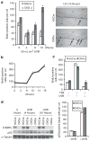
Figure 5. C/EBPα (CCAAT/enhancer-binding protein-α) has a role in UVB-induced inhibition of cell cycle progression in vivo
BrdU was injected 1 hour before killing. Data are expressed as BrdU-positive cells per cm length of skin±SE. N≥3/genotype/treatment. (a) Mice were treated with UVB. #Different from untreated mice of the same genotype, P<0.05; *not different from untreated mice of the same genotype, P>0.05. Photomicrographs of BrdU staining. Scale bar=10 μm. (b) TPA (12-O-tetradecanoylphorbol-13-acetate) treatment causes keratinocytes to enter S phase. (c) Mice were treated with TPA and 4 hours later, mice were either treated with UVB or not and they were killed at 14 hours post-TPA. *Different CKOα TPA/UVB versus keratin 5 Cre (K5Cre) TPA/UVB, P<0.05. (d) Immunoblot analysis of epidermal lysates before and after UVB treatment. The right panel shows p53 immunohistochemical (IHC)-positive basal keratinocytes in mouse skin±UVB.

References

    1. American Cancer Society. Cancer Facts & Figures 2008. 2008 http://www.cancer.org/Research/CancerFactsFigures/cancer-facts-figures-2008.
    1. Benavides F, Oberyszyn TM, VanBuskirk AM, et al. The hairless mouse in skin research. J Dermatol Sci. 2009;53:10–8. - PMC - PubMed
    1. Bennett KL, Hackanson B, Smith LT, et al. Tumor suppressor activity of CCAAT/e. Cancer Res. 2007;67:4657–64. - PubMed
    1. Bielas JH, Loeb KR, Rubin BP, et al. Human cancers express a mutator phenotype. Proc Natl Acad Sci USA. 2006;103:18238–42. - PMC - PubMed
    1. Bielas JH, Loeb LA. Quantification of random genomic mutations. Nat Methods. 2005;2:285–90. - PubMed

Publication types

MeSH terms

Substances