. 2011 Jun;131(6):1272-9.
doi: 10.1038/jid.2011.24. Epub 2011 Feb 24.

TNF-α downregulates filaggrin and loricrin through c-Jun N-terminal kinase: role for TNF-α antagonists to improve skin barrier

Affiliations

TNF-α downregulates filaggrin and loricrin through c-Jun N-terminal kinase: role for TNF-α antagonists to improve skin barrier

Byung Eui Kim et al. J Invest Dermatol. 2011 Jun.

Erratum in

  • J Invest Dermatol. 2011 Jun;131(6):1388. Guttman, Emma [corrected to Guttman-Yassky, Emma]

Abstract

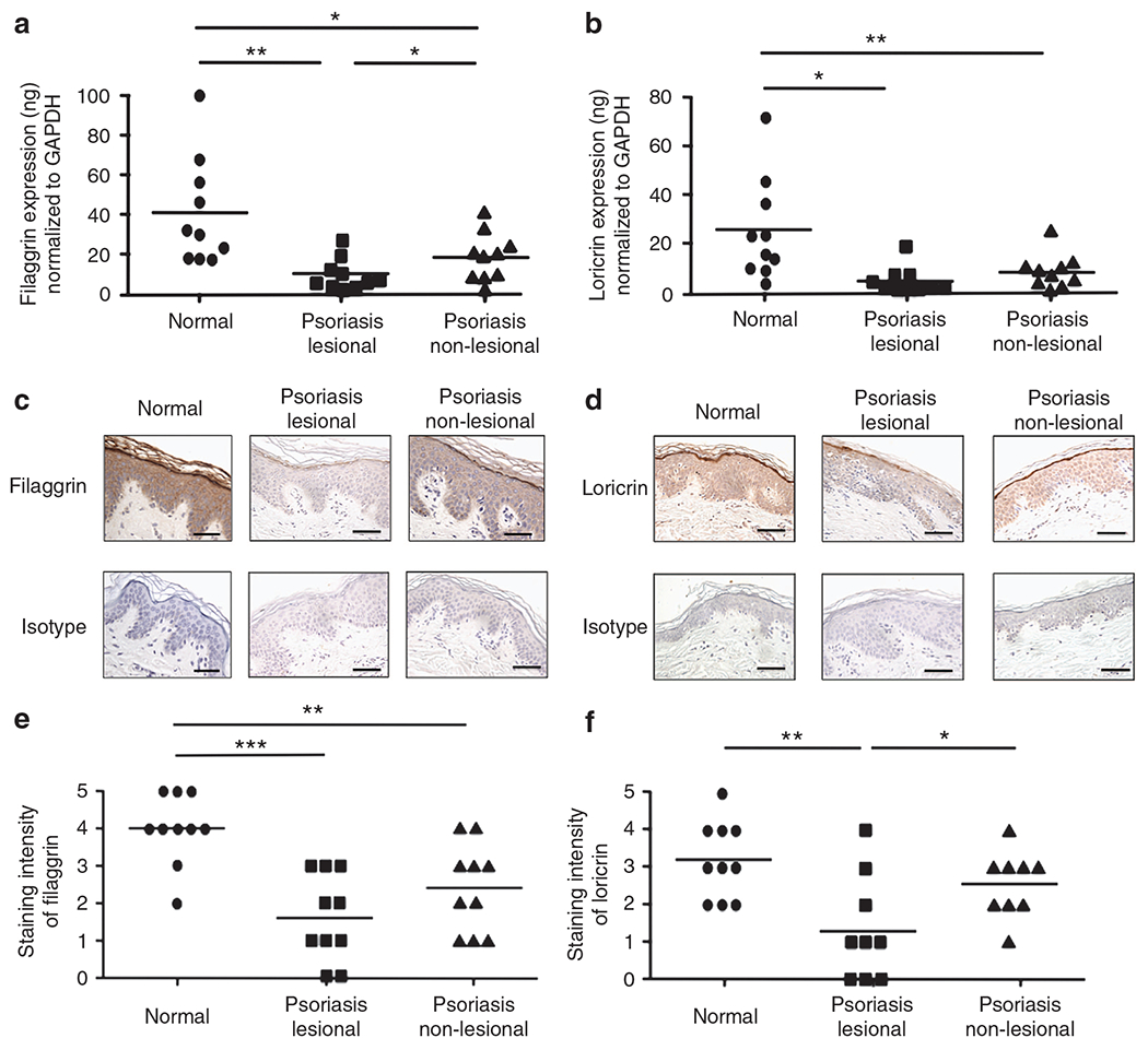
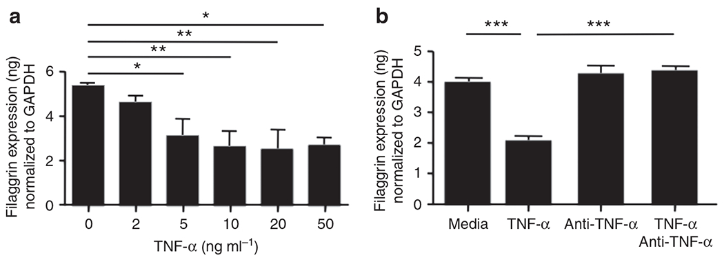
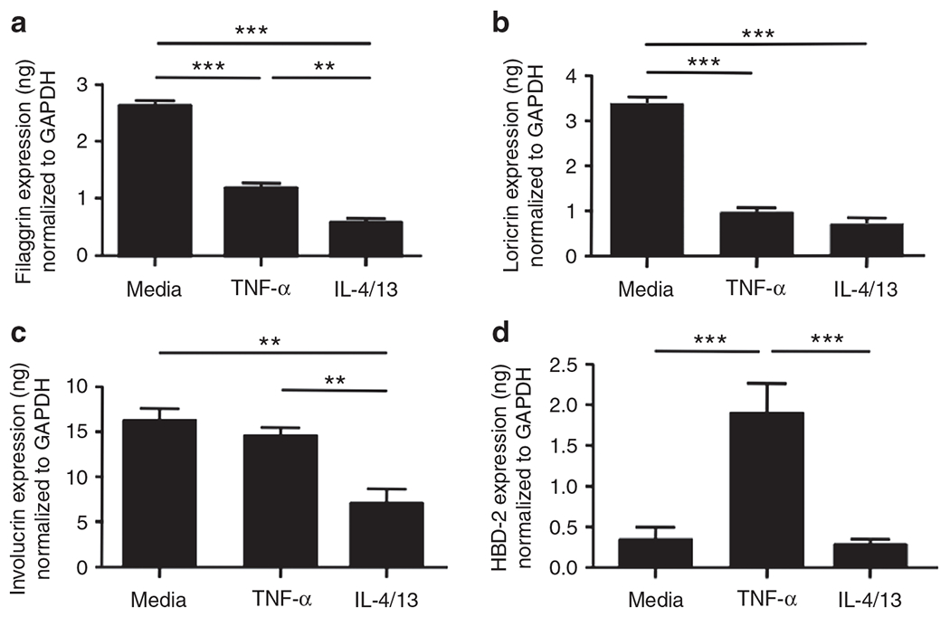
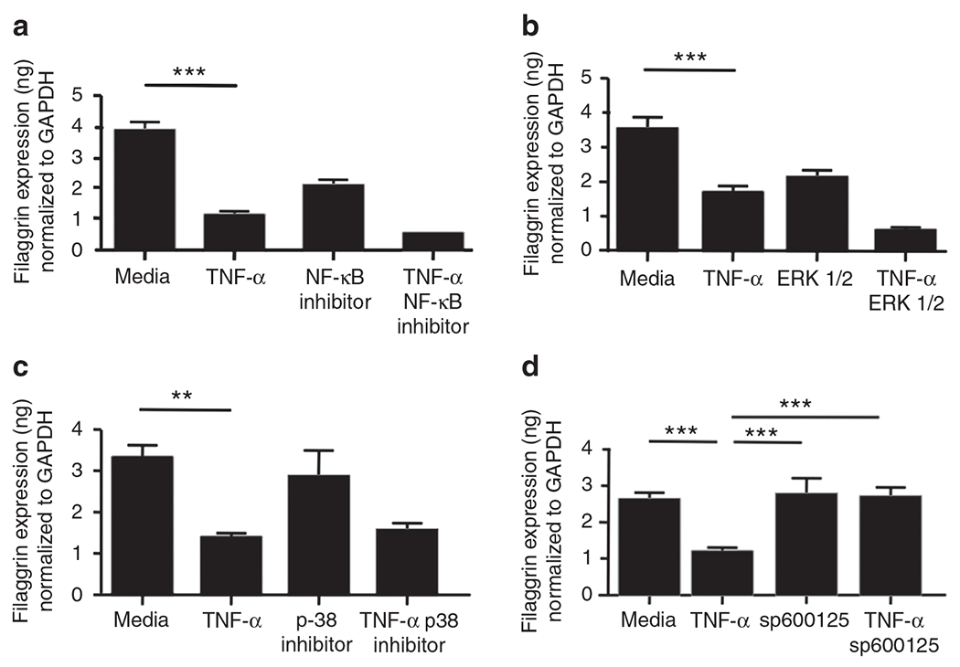
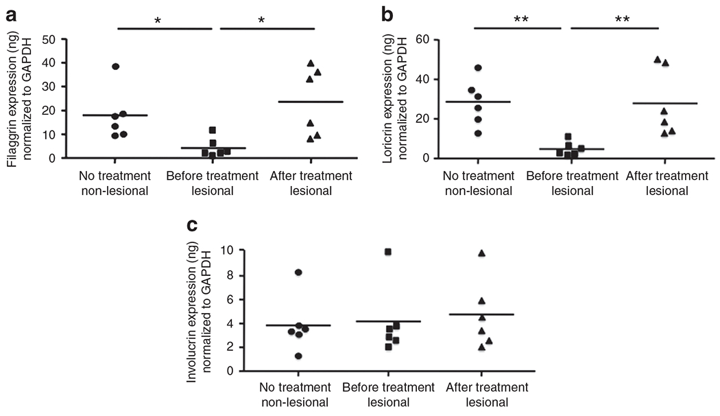
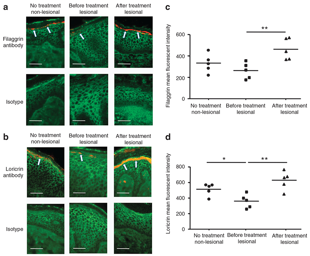
Filaggrin (FLG), loricrin (LOR), and involucrin are important epidermal barrier proteins. As psoriasis is characterized by overexpression of tumor necrosis factor-α (TNF-α) and impaired skin barrier, we investigated the expression of skin barrier proteins in psoriasis patients and whether their expression was modulated by TNF-α. The expression of FLG and LOR was found to be decreased in lesional and non-lesional skin of psoriasis patients. A correlation was found between the expression of TNF-α and epidermal barrier proteins in psoriasis. TNF-α was found to modulate the expression of FLG and LOR via a c-Jun N-terminal kinase-dependent pathway. Importantly, we report that clinical treatment of psoriasis patients with a TNF-α antagonist results in significant enhancement of epidermal barrier protein expression. Our current study suggests that TNF inhibits barrier protein expression, and TNF-α antagonists may contribute to clinical improvement in patients with psoriasis by improving barrier protein expression.


Conflict of interest statement

CONFLICT OF INTEREST

The authors state no conflict of interest.

Figures

Figure 1.
Figure 1.. Expression of filaggrin (FLG) and loricrin (LOR) in the skin from normal subjects and patients with psoriasis.
RNA was isolated from the skin of normal subjects and patients with psoriasis. The gene expression FLG (a) and LOR (b) was evaluated using real-time RT-PCR. (c, d) Representative paraffin-embedded skin biopsies from normal subjects (n = 10) and patients with psoriasis (n = 9) stained for FLG (c) and LOR (d) are shown. Images were collected at × 400 magnification. (e, f) The intensities of the staining for FLG (e) and LOR (f) were graded visually on a scale from 0 (no staining) to 5 (the most intense staining). The scale bar represents 50 μm. *P<0.05; **P<0.01; ***P<0.001. GAPDH, glyceraldehyde-3-phosphate dehydrogenase.
Figure 2.
Figure 2.. Tumor necrosis factor-α (TNF-α) inhibits expression of filaggrin (FLG).
Primary human keratinocytes (KCs) were stimulated in the presence of various concentrations of TNF-α for 24 hours. The gene expression of FLG was examined by real-time RT-PCR (a). In addition, primary human KCs were pre-incubated with 0.1 μg ml−1 of TNF-α-neutralizing antibody before TNF-α (20 ng ml−1) stimulation. The gene expression of FLG was then examined by real-time RT-PCR (b). *P<0.05; **P<0.01; ***P<0.001. GAPDH, glyceraldehyde-3-phosphate dehydrogenase.
Figure 3.
Figure 3.. The effects of tumor necrosis factor-α (TNF-α) and Th2 cytokines on barrier proteins.
Primary human keratinocytes were stimulated with TNF-α (20 ng ml−1) or a combination of IL-4 (50 ng ml−1) and IL-13 (50 ng ml−1) for 24 hours. The gene expression of filaggrin (FLG) (a), loricrin (LOR) (b), involucrin (IVL) (c), and human-β defensin 2 (HBD-2) (d) were examined by real-time RT-PCR. **P<0.01; ***P<0.001. GAPDH, glyceraldehyde-3-phosphate dehydrogenase.
Figure 4.
Figure 4.. c-Jun N-terminal kinase (JNK) inhibitor blocks the tumor necrosis factor-α (TNF-α)-mediated inhibition of filaggrin (FLG).
Primary human keratinocytes were pre-incubated with NF-κB inhibitor (10 nm, a), extracellular signal-regulated kinases 1/2 (ERK1/2) inhibitor (5 μm, b), p38 inhibitor (5 μm, c), and JNK inhibitor sp600125 (1 μm, d) before stimulation with TNF-α (20 ng ml−1). The gene expression of FLG was examined by real-time RT-PCR. **P<0.01; ***P<0.001. GAPDH, glyceraldehyde-3-phosphate dehydrogenase.
Figure 5.
Figure 5.. Tumor necrosis factor-α (TNF-α) antagonist increased the gene expression of filaggrin (FLG), loricrin (LOR), and involucrin (IVL).
Patients with psoriasis (n = 6) were treated with TNF-α monoclonal antibody for 12 weeks. RNA was isolated from the lesional and non-lesional skin of the patients with psoriasis. Gene expression of FLG (a), LOR (b), and IVL (c) was evaluated using real-time RT-PCR. *P<0.05, **P<0.01. GAPDH, glyceraldehyde-3-phosphate dehydrogenase.
Figure 6.
Figure 6.. Tumor necrosis factor-α (TNF-α) antagonist increased the protein expression of filaggrin (FLG) and loricrin (LOR).
(a, b) Representative skin biopsies from patients with psoriasis stained for FLG (a) and LOR (b) are shown. (c, d) The mean fluorescent intensity is shown for FLG (c) and LOR (d) in the epidermis of each biopsy. The scale bar represents 50 μm. *P<0.05, **P<0.01. Arrows in panel a and b indicate protein expression of filaggrin and loricrin, respectively.

Similar articles

Cited by

References

    1. Albanesi C, Fairchild HR, Madonna S et al. (2007) IL-4 and IL-13 negatively regulate TNF-alpha- and IFN-gamma-induced beta-defensin expression through STAT-6, suppressor of cytokine signaling (SOCS)-1, and SOCS-3. J Immunol 179:984–92 - PubMed
    1. Candi E, Schmidt R, Melino G (2005) The cornified envelope: a model of cell death in the skin. Nat Rev Mol Cell Biol 6:328–40 - PubMed
    1. Chang L, Karin M (2001) Mammalian MAP kinase signalling cascades. Nature 410:37–40 - PubMed
    1. Chiricozzi A, Guttman-Yassky E, Suárez-Fariñas M et al. (2010) Integrative responses to IL-17 and TNF-alpha in human keratinocytes account for key inflammatory pathogenic circuits in psoriasis. J Invest Dermatol 131:677–87 - PubMed
    1. Christophers E, Henseler T (1987) Contrasting disease patterns in psoriasis and atopic dermatitis. Arch Dermatol Res 279(Suppl):S48–51 - PubMed

Publication types

MeSH terms