. 2011 Feb 8;9(2):e1000590.
doi: 10.1371/journal.pbio.1000590.

Noncanonical compensation of zygotic X transcription in early Drosophila melanogaster development revealed through single-embryo RNA-seq

Affiliations

Noncanonical compensation of zygotic X transcription in early Drosophila melanogaster development revealed through single-embryo RNA-seq

Susan E Lott et al. PLoS Biol. .

Abstract

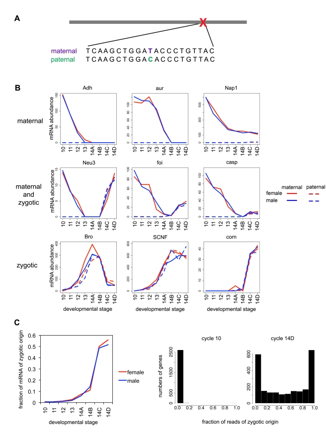
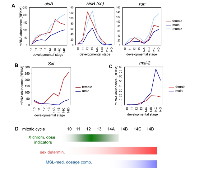
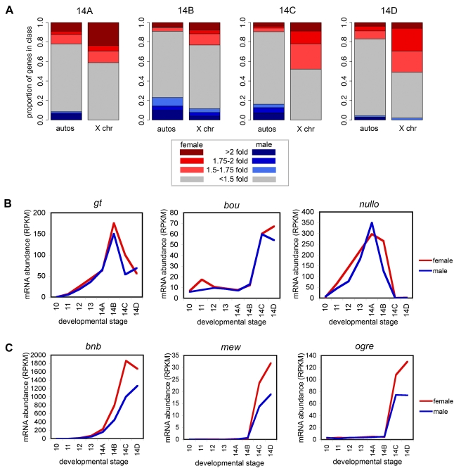
When Drosophila melanogaster embryos initiate zygotic transcription around mitotic cycle 10, the dose-sensitive expression of specialized genes on the X chromosome triggers a sex-determination cascade that, among other things, compensates for differences in sex chromosome dose by hypertranscribing the single X chromosome in males. However, there is an approximately 1 hour delay between the onset of zygotic transcription and the establishment of canonical dosage compensation near the end of mitotic cycle 14. During this time, zygotic transcription drives segmentation, cellularization, and other important developmental events. Since many of the genes involved in these processes are on the X chromosome, we wondered whether they are transcribed at higher levels in females and whether this might lead to sex-specific early embryonic patterning. To investigate this possibility, we developed methods to precisely stage, sex, and characterize the transcriptomes of individual embryos. We measured genome-wide mRNA abundance in male and female embryos at eight timepoints, spanning mitotic cycle 10 through late cycle 14, using polymorphisms between parental lines to distinguish maternal and zygotic transcription. We found limited sex-specific zygotic transcription, with a weak tendency for genes on the X to be expressed at higher levels in females. However, transcripts derived from the single X chromosome in males were more abundant that those derived from either X chromosome in females, demonstrating that there is widespread dosage compensation prior to the activation of the canonical MSL-mediated dosage compensation system. Crucially, this new system of early zygotic dosage compensation results in nearly identical transcript levels for key X-linked developmental regulators, including giant (gt), brinker (brk), buttonhead (btd), and short gastrulation (sog), in male and female embryos.


Conflict of interest statement

MBE is a cofounder and member of the Board of Directors of PLoS.

Figures

Figure 1
Figure 1. A sex-specific timecourse of early-embryonic gene expression.
(A) Transcription events during early embryogenesis. During the first 8–9 mitotic cycles, almost all RNAs in the embryo are of maternal origin. Zygotic transcription begins at a low level at approximately cycle 10 and becomes widespread by the middle of cycle 14. MSL-mediated dosage compensation begins late in or following cycle 14. (B) Embryos used for mRNA-Seq. Individual embryos in the interphases of cycles 10 to 14 were selected by direct observation of mitosis in embryos containing histone H2Av-RFP and computing nuclear density. Embryos at substages of cycle 14 were selected by observing the extent of progression through cellularization (from proportion of membrane invagination) under light microscopy. Each embryo pictured here was placed into TRIzol reagent immediately after these images were taken, DNA and RNA were extracted, and each sample was genotyped to determine the sex of the embryo. (C) Approximately 100 ng total RNA was obtained from each embryo, and poly-A RNA was processed with an amplification-free protocol optimized for small samples and sequenced on an Illumina GAIIx Genome Analyzer. Data (normalized reads per kb, RPKM) from independently processed individuals of the same stage and same sex, and same stage but different sex were extremely similar, while individuals from different stages showed larger numbers of differences.
Figure 2
Figure 2. Polymorphisms distinguish maternal and zygotic expression.
(A) Approximately 70% of genes expressed in the early embryo contained fixed differences between the maternal (w1) and paternal (CantonS) lines, allowing us to partition the expression level for that gene at each time point into those derived from the maternal and paternal chromosomes. (B) We classified genes based on temporal profiles of total mRNA and (where available) mRNA derived from maternal and paternal chromosomes. Maternally deposited transcripts (∼5,000) were expressed at high levels that decay over time and come exclusively from the maternal chromosome. Zygotic transcripts (∼2,000) were not present or were present at very low levels at cycle 10, and transcript levels rose over time with equal contribution from maternal and paternal chromosomes. Approximately 800 transcripts are both maternally deposited and zygotically transcribed. (C) Left, the average proportion of zygotic reads per gene increases over time, accelerating during mid cycle 14. Right, a histogram showing the proportion of zygotic reads over genes for an early and a late stage.
Figure 3
Figure 3. Transcription of sex determination and dosage compensation genes.
The events in the sex determination pathway in our data are consistent with previous studies. (A) Expression levels (normalized reads per kb, RPKM) for the X chromosome signal elements (XSEs; sisA, sisB, and run) in female embryos reach twice the transcript abundance of male embryos (light blue line) near cycle 12. These factors activate Sxl expression (B) in females, with significant female expression levels around cycle 13, the presence of which interferes with msl-2 expression (C), the male-specific protein of the MSL-mediated dosage compensation, which is higher in males, starting mid-late cycle 14.
Figure 4
Figure 4. Zygotic transcription from the X chromosome is weakly female biased.
(A) Female expression versus male expression for zygotic genes (normalized reads per kb, per gene, log scale) over cycle 14, where most zygotic expression is detected. Autosomal gene expression was centered around the purple line, where female and male transcript levels are even. For X chromosomal genes, transcript levels were distributed between females and males having equal transcript abundance (solid line) and the female having twice the transcript level of the male (dotted line). (B) Total expression levels (average normalized reads per gene) for zygotic genes in male and female embryos, on autosomes and the X chromosome. Female expression on X is less than twice the level of male expression after cycle 12 (light blue line). (C) Zygotic transcripts from autosomal genes were derived equally from the maternal or paternal chromosomes, while zygotic transcripts from the single X chromosome in males are present at higher levels than those from either of the X chromosomes in females, demonstrating that the early embryo is dosage compensated.
Figure 5
Figure 5. Early zygotic dosage compensation.
Of the zygotic genes on the X chromosome, some had the same transcript levels in female and male embryos, and some had an excess of transcripts in females relative to the male, indicating that some are transcriptionally dosage compensated and some are not. (A) Proportion of genes that had higher transcript levels in males or females over cycle 14, comparing autosomes to the X chromosome. The darker colors represent a stronger enrichment of female or male transcripts relative to the other sex. To reduce noise, ratios of female to male expression were considered for genes where individuals of both sexes had at least 2 RPKM, little qualitative difference was observed in results for higher thresholds (results not shown). (B) Key developmental regulators on the X chromosome were dosage compensated at the transcript level. (C) Other zygotic factors on the X did not appear to be effectively dosage compensated, as there were large differences in expression between male and female embryos.

Comment in

  • Doubling the dose.
    Mair W. Mair W. PLoS Biol. 2011 Feb 8;9(2):e1001017. doi: 10.1371/journal.pbio.1001017. PLoS Biol. 2011. PMID: 21346808 Free PMC article. No abstract available.

References

    1. Pritchard D. K, Schubiger G. Activation of transcription in Drosophila embryos is a gradual process mediated by the nucleocytoplasmic ratio. Genes Dev. 1996;10:1131–1142. - PubMed
    1. Cline T. W, Meyer B. J. Vive la difference: males vs females in flies vs worms. Annu Rev Genet. 1996;30:637–702. - PubMed
    1. Schutt C, Nothiger R. Structure, function and evolution of sex-determining systems in Dipteran insects. Development. 2000;127:667–677. - PubMed
    1. Erickson J. W, Quintero J. J. Indirect effects of ploidy suggest X chromosome dose, not the X∶A ratio, signals sex in Drosophila. PLoS Biol. 2007;5:e332. doi: 10.1371/journal.pbio.0050332. - DOI - PMC - PubMed
    1. Gelbart M. E, Kuroda M. I. Drosophila dosage compensation: a complex voyage to the X chromosome. Development. 2009;136:1399–1410. - PMC - PubMed

Publication types

MeSH terms