

Mitochondrial disorders with significant ophthalmic manifestations
- PMID: 21346843
- PMCID: PMC3038114
- DOI: 10.4103/0974-9233.51998
Mitochondrial disorders with significant ophthalmic manifestations
Abstract
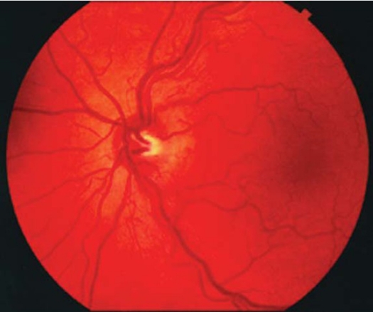
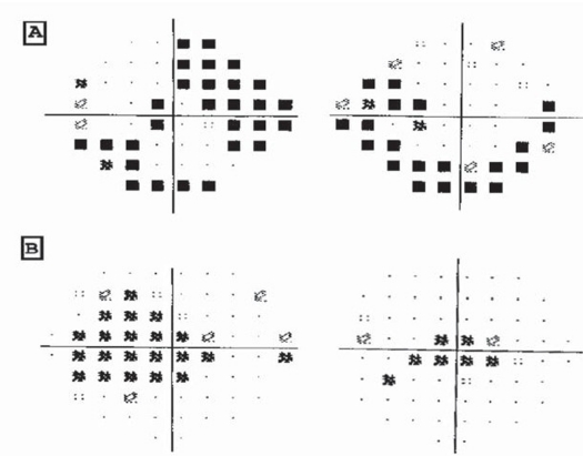
Mitochondrial diseases are a clinically hetyerogenous group of disorders. They can be caused by mutations of nuclear or mitochondrial DNA (mtDNA). Some affect a single organ, but many involve multiple organ systems and often present with prominent neurologic and myopathic features. The eye is frequently affected, along with muscles and brain, but multisystem disease is common. Ophthalmic manifestations include cataract, retinopathy, optic atrophy, cortical visual loss, ptosis and ophthalmoplegia. Kearns-Sayre Syndrome (KSS), Mitochondrial Encephalopathy, Lactic Acidosis Stroke (MELAS), Myoclonic Epilepsy and Ragged Red Fiber myopathy (MERRF) and Lebers Hereditary Optic Neuropathy (LHON) are well known clinical entities that are secondary to mtDNA abnormalities, which has ophthalmic manifestations. Mitochondrial Dysfunction should be considered in the differential diagnosis of progressive multisystem disorder and specifically if there is associated neuro-ophthalmic manifestations, which may be the presenting symptom of these disorders.
Keywords: Mitochondrial disorder genetics; diagnosis; variable manifestations.
Figures
References
-
- Di Mauro S, Shon EA. Mitochondrial respiratory – chain disease. The New England Journal of Medicine. 2003;348:2656–2668. - PubMed
-
- Finsterer J. Central nervous system manifestations of mitochondrial disorders. Acta Neruol Second. 2006;114:217–238. - PubMed
-
- Bionsse Neuro-ophthalmology of mitochondrial diseases. Carr Opin Neural. 2003;16(1):35–43. - PubMed
-
- Chinnery PF. Mitochondrial disorders overview. Gene Review. 2006. Feb, pp. 1–20.
-
- Newman NJ. Hereditary optic neuropathies: from the mitochondria to the optic nerve. American Journal of Ophthalmology. 2005;140:517–523. - PubMed
LinkOut - more resources
Full Text Sources
