

Evolution of an Astrocytic Hamartoma of the Optic Nerve Head in a Patient with Retinitis Pigmentosa - Photographic Documentation over 2 Years of Follow-Up
- PMID: 21347192
- PMCID: PMC3042018
- DOI: 10.1159/000324037
Evolution of an Astrocytic Hamartoma of the Optic Nerve Head in a Patient with Retinitis Pigmentosa - Photographic Documentation over 2 Years of Follow-Up
Abstract
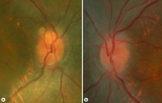
Aim: To report photographically the evolution of an astrocytic hamartoma of the left optic nerve head over a 2-year follow-up in a patient with retinitis pigmentosa.
Methods: A 14-year-old boy was seen in the medical retina clinic with a 3-year history of night blindness. Best corrected visual acuity was 6/18 in both eyes. Colour vision was normal in both eyes and confrontation fields showed peripheral constriction. Fundus examination revealed bone spicule pigmentary changes at the retinal mid periphery typical of retinitis pigmentosa and superficial globules at the margins of both optic nerve heads. Electrodiagnostic tests confirmed moderately severe rod cone dystrophy with macular involvement bilaterally.
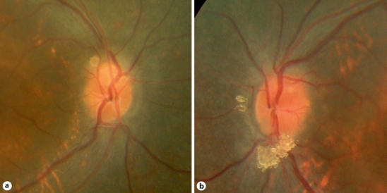
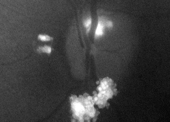
Results: Two years later, the ocular examination was unchanged except for the appearance of the optic nerve head lesion in the left eye. There was an increase in the size of the lesion which was diagnosed as an astrocytic hamartoma. Further investigations were recommended to exclude neurofibromatosis and tuberous sclerosis.
Conclusion: Astrocytic hamartomas of the optic nerve head and optic nerve head drusen have both been described in patients with retinitis pigmentosa. They can be a diagnostic dilemma although drusen are more common (10%). To differentiate these two entities it is very important to document any growth during the follow-up period which is suggestive of astrocytic hamartoma rather than optic disc drusen.
Keywords: Astrocytic hamartoma; Optic disc drusen; Retinitis pigmentosa.
Figures
References
-
- Williams R, Taylor D. Tuberous sclerosis. Surv Ophthalmol. 1985;30:143–154. - PubMed
-
- Ulbright TM, Fulling KH, Helveston EM. Astrocytic tumours of the retina. Differentiation of sporadic tumours from phakomatoses-associated tumours. Arch Pathol Lab Med. 1984;108:160–163. - PubMed
-
- Reeser FH, Aaberg TM, Van Horn DL. Astrocytic hamartoma of the retina not associated with tuberous sclerosis. Am J Ophthalmol. 1978;86:688–698. - PubMed
-
- Bec P, Mathis A, Adam P, Maillard P, Alberge Y. Retinitis pigmentosa associated with astrocytic hamartomas of the optic disc. Ophthalmologica. 1984;189:135–138. - PubMed
-
- Pillai S, Limaye S, Saimovici LB. Optic disc hamartoma associated with retinitis pigmentosa. Retina. 1983;3:24–26.
Publication types
LinkOut - more resources
Full Text Sources
Research Materials
