. 2011 Feb 11;6(2):e17119.
doi: 10.1371/journal.pone.0017119.

Unusual structures are present in DNA fragments containing super-long Huntingtin CAG repeats

Affiliations

Unusual structures are present in DNA fragments containing super-long Huntingtin CAG repeats

Daniel Duzdevich et al. PLoS One. .

Abstract

Background: In the R6/2 mouse model of Huntington's disease (HD), expansion of the CAG trinucleotide repeat length beyond about 300 repeats induces a novel phenotype associated with a reduction in transcription of the transgene.

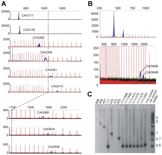
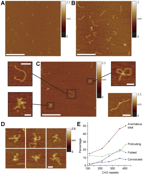
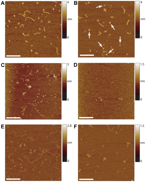
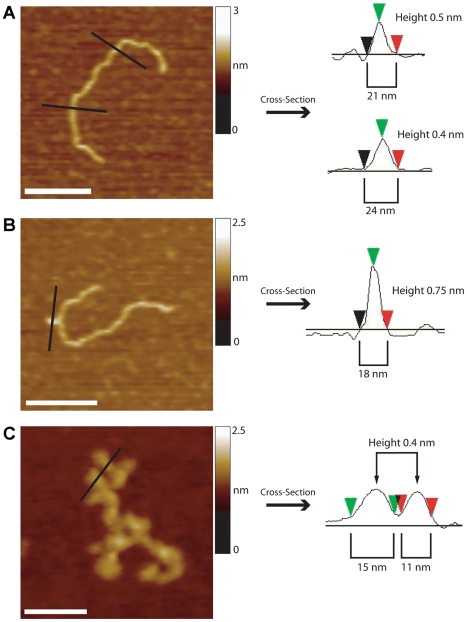
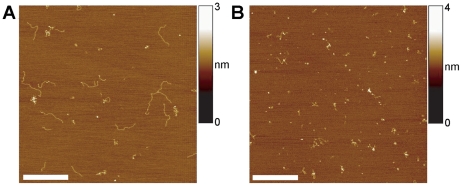
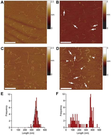
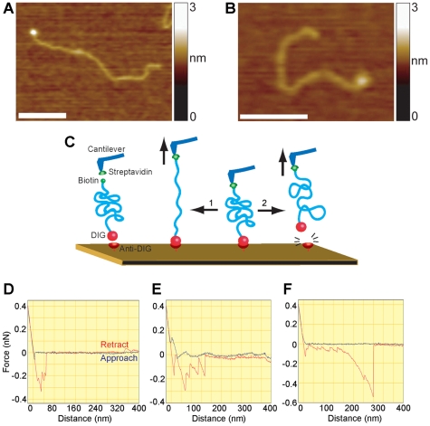
Methodology/principal findings: We analysed the structure of polymerase chain reaction (PCR)-generated DNA containing up to 585 CAG repeats using atomic force microscopy (AFM). As the number of CAG repeats increased, an increasing proportion of the DNA molecules exhibited unusual structural features, including convolutions and multiple protrusions. At least some of these features are hairpin loops, as judged by cross-sectional analysis and sensitivity to cleavage by mung bean nuclease. Single-molecule force measurements showed that the convoluted DNA was very resistant to untangling. In vitro replication by PCR was markedly reduced, and TseI restriction enzyme digestion was also hindered by the abnormal DNA structures. However, significantly, the DNA gained sensitivity to cleavage by the Type III restriction-modification enzyme, EcoP15I.

Conclusions/significance: "Super-long" CAG repeats are found in a number of neurological diseases and may also appear through CAG repeat instability. We suggest that unusual DNA structures associated with super-long CAG repeats decrease transcriptional efficiency in vitro. We also raise the possibility that if these structures occur in vivo, they may play a role in the aetiology of CAG repeat diseases such as HD.


Conflict of interest statement

Competing Interests: JJ and JW are employees of Laragen, Inc., a DNA sequencing company. This does not alter the authors' adherence to all the PLoS ONE policies on sharing data and materials.

Figures

Figure 1
Figure 1. CAG repeat length affects PCR efficiency and accuracy.
(A) GeneScan profiles of PCR products containing various CAG repeat lengths, showing that peak heights fall and peak widths rise as CAG repeat length rises. (Note the changing values on the y-axes). The red peaks indicate the positions of size markers (bp). A bp-scale is shown at the top. (B) GeneScan profiles of PCR products from two double mutant mice, showing that in both cases the longer repeat is under-represented. (C) Agarose gel of PCR products containing various repeat lengths. CAG repeat lengths are indicated above each lane. The left-hand side shows PCR products generated from DNA taken from mice carrying single copy of the transgene of varying sizes; the right-hand side shows PCR products from reactions containing equal amounts of the DNA from two of the mice shown on the left hand side; one carrying a CAG repeat of 111 and one other carrying a longer repeat. Note that the PCR reaction in the lane labelled 373 has failed.
Figure 2
Figure 2. Unusual structures occur in super-long CAG repeat DNA.
(A) AFM image of wild type DNA with 8 CAG repeats (total length 298 bp). Scale bar, 500 nm. A colour-height scale is shown at the right. (B) DNA with 216 repeats (total length 779 bp). Scale bar, 500 nm. (C) DNA with 360 CAG repeats (total length 1211 bp). Scale bar, 500 nm. DNA fragments were classified into ‘normal and linear’ (inset, upper left; scale bar 100 nm), ‘convoluted’ (lower left; scale bar, 50 nm), ‘folded’ (upper right; scale bar, 50 nm), or ‘protruding’ (lower right, derived from another image of the same PCR product; scale bar 100 nm). (D) Gallery of zoomed images showing examples of convoluted DNA. (E) Graph showing the relationship between the percentage of DNA molecules with unusual structures and CAG repeat length. Numbers of DNA molecules analysed ranged from 1163 (114 repeats) to 203 (408 repeats).
Figure 3
Figure 3. Convoluted DNA does not contain extensive regions of ssDNA.
(A) AFM image of human σ-1 receptor DNA, generated by PCR using a 50-fold excess of forward primer. Two types of structure can be seen: 690-bp dsDNA, which is linear and ‘normal’, and ssDNA, which is very dense and compacted. Scale bar, 250 nm. (B) σ-1 receptor DNA after extensive Tse1 digestion at 80°C. The enzyme has cleaved the dsDNA, leaving shorter fragments, but the ssDNA (arrows) remains unaffected. Scale bar, 250 nm. (C) DNA containing 585 CAG repeats. Note that some molecules are linear, whereas others are folded or convoluted. Scale bar, 500 nm. (D) 585-repeat DNA after extensive Tse1 digestion at 80°C. A dense homogenous ‘background’ of short (∼6-bp) fragments remains, together with a few aggregations of these fragments, and one fragment of ∼60 bp, which likely represents a flanking region. Scale bar, 250 nm. (E) Undigested 585-repeat DNA. Scale bar, 500 nm. (F) 560-repeat DNA after limited Tse1 digestion at room temperature. The enzyme has preferentially cleaved linear regions of DNA, leaving behind the anomalous regions. Scale bar, 250 nm.
Figure 4
Figure 4. Sections through DNA molecules showing either normal or unusual structures.
(A) Normal DNA (216 CAG repeats). Scale bar, 100 nm. (B) Protruding DNA (360 CAG repeats). The cross-section shows a width comparable to that of normal dsDNA but a greater height, suggesting an imperfect hairpin or hairpin-like structure. Scale bar, 100 nm. (C) Convoluted DNA (585 CAG repeats). The cross-section indicates protrusions similar in height and width to dsDNA. Scale bars, 50 nm.
Figure 5
Figure 5. Effect of mung bean nuclease indicates the presence of hairpins in the convoluted molecules.
(A) DNA containing 360 CAG repeats that had been treated with mung bean nuclease. Note the presence of an unusual number of short fragments. (B) The same DNA that had been treated with mung bean nuclease, and then melted and reannealed. Note the almost complete absence of full-length linear DNA. Scale bars, 250 nm.
Figure 6
Figure 6. Convoluted DNA is selectively digested by EcoP15I.
(A, B) AFM images of DNA with 216 CAG repeats (total length 740 bp) before (A) and after (B) incubation with EcoP15I. (C, D) DNA with 360 CAG repeats (total length 1211 bp) before (C) and after (D) incubation with EcoP15I. Arrows in (D) indicate DNA molecules bearing enzyme bound at the ends. Arrowheads indicate residual tightly-wound tangles. All scale bars, 500 nm. (E, F) Frequency distributions of lengths of linear DNA in untreated (E) and EcoP15I-treated (F) samples of 360-repeat DNA.
Figure 7
Figure 7. Convoluted structures resist considerable pulling force.
(A) AFM image of a DNA molecule containing 360 CAG repeats (total length 1211 bp) showing a streptavidin molecule, attached to a terminal biotin moiety. Scale bar, 100 nm. (B) AFM image of a 272-repeat DNA molecule showing an anti-DIG antibody molecule, attached to a terminal DIG moiety. Scale bar, 100 nm. (C) Experimental rationale. A silicon nitride cantilever and a mica disk were both amino-functionalized by incubation with APTES. Glutaraldehyde was then used to couple streptavidin to the cantilever and anti-DIG to the mica. DNA fragments were bound to the steptavidin via a biotin tag on the DNA. The DNA was lowered onto the antibody-coated mica to allow the DIG tag on the DNA to bind to its antibody on the mica. The cantilever was then retracted, and the forces applied during the retraction are measured. Two possible outcomes are depicted: (1) the convoluted DNA is unwound; (2) the DNA remains convoluted and the antigen-antibody bond is ruptured. (D) Typical force curve for a DNA fragment containing 114 CAG repeats, showing ‘approach’ (blue) and ‘retract’ (red) traces. Note the multiple force peaks, the final rupture distance (41 nm) and force (200 pN). 14/150 retractions (9%) resulted in a measurable force peak. (E) Typical force curve for a DNA fragment containing 360 CAG repeats. Note the multiple force peaks, the final rupture distance (117 nm) and force (159 pN). 22/124 retractions (18%) resulted in a measurable force peak. (F) Force curve for a 360-repeat fragment showing a very long rupture distance (260 nm) and a large force (529 pN).

Similar articles

Cited by

References

    1. Orr HT, Zoghbi HY. Trinucleotide repeat disorders. Annu Rev Neurosci. 2007;30:575–621. - PubMed
    1. López Castel A, Cleary JD, Pearson CE. Repeat instability as the basis for human diseases and as a potential target for therapy. Nat Rev Mol Cell Biol. 2010;11:165–170. - PubMed
    1. Wexler NS, Lorimer J, Porter J, Gomez F, Moskowitz C, et al. Venezuelan kindreds reveal that genetic and environmental factors modulate Huntington's disease age of onset. Proc Natl Acad Sci USA. 2004;101:3498–3503. - PMC - PubMed
    1. Li JL, Hayden MR, Warby SC, Durr A, Morrison PJ, et al. Genome-wide significance for a modifier of age at neurological onset in Huntington's disease at 6q23-24: the HD MAPS study. BMC Med Genet. 2006;7:71. - PMC - PubMed
    1. Andresen JM, Gayán J, Djousse L, Roberts S, Brocklebank D, et al. The relationship between CAG repeat length and age of onset differs for Huntington's disease patients with juvenile onset or adult onset. Ann Hum Genet. 2007;71:295–301. - PubMed

Publication types

MeSH terms