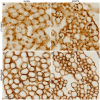
Phosphoinositide regulation of integrin trafficking required for muscle attachment and maintenance
- PMID: 21347281
- PMCID: PMC3037412
- DOI: 10.1371/journal.pgen.1001295
Phosphoinositide regulation of integrin trafficking required for muscle attachment and maintenance
Abstract
Muscles must maintain cell compartmentalization when remodeled during development and use. How spatially restricted adhesions are regulated with muscle remodeling is largely unexplored. We show that the myotubularin (mtm) phosphoinositide phosphatase is required for integrin-mediated myofiber attachments in Drosophila melanogaster, and that mtm-depleted myofibers exhibit hallmarks of human XLMTM myopathy. Depletion of mtm leads to increased integrin turnover at the sarcolemma and an accumulation of integrin with PI(3)P on endosomal-related membrane inclusions, indicating a role for Mtm phosphatase activity in endocytic trafficking. The depletion of Class II, but not Class III, PI3-kinase rescued mtm-dependent defects, identifying an important pathway that regulates integrin recycling. Importantly, similar integrin localization defects found in human XLMTM myofibers signify conserved MTM1 function in muscle membrane trafficking. Our results indicate that regulation of distinct phosphoinositide pools plays a central role in maintaining cell compartmentalization and attachments during muscle remodeling, and they suggest involvement of Class II PI3-kinase in MTM-related disease.
Conflict of interest statement
The authors have declared that no competing interests exist.
Figures


Similar articles
-
Drosophila Mtm and class II PI3K coregulate a PI(3)P pool with cortical and endolysosomal functions.J Cell Biol. 2010 Aug 9;190(3):407-25. doi: 10.1083/jcb.200911020. J Cell Biol. 2010. PMID: 20696708 Free PMC article.
-
AAV-mediated intramuscular delivery of myotubularin corrects the myotubular myopathy phenotype in targeted murine muscle and suggests a function in plasma membrane homeostasis.Hum Mol Genet. 2008 Jul 15;17(14):2132-43. doi: 10.1093/hmg/ddn112. Epub 2008 Apr 22. Hum Mol Genet. 2008. PMID: 18434328 Free PMC article.
-
PIK3C2B inhibition improves function and prolongs survival in myotubular myopathy animal models.J Clin Invest. 2016 Sep 1;126(9):3613-25. doi: 10.1172/JCI86841. Epub 2016 Aug 22. J Clin Invest. 2016. PMID: 27548528 Free PMC article.
-
Implication of phosphoinositide phosphatases in genetic diseases: the case of myotubularin.Cell Mol Life Sci. 2003 Oct;60(10):2084-99. doi: 10.1007/s00018-003-3062-3. Cell Mol Life Sci. 2003. PMID: 14618257 Free PMC article. Review.
-
Myotubularin phosphoinositide phosphatases in human diseases.Curr Top Microbiol Immunol. 2012;362:209-33. doi: 10.1007/978-94-007-5025-8_10. Curr Top Microbiol Immunol. 2012. PMID: 23086420 Review.
Cited by
-
SPEG binds with desmin and its deficiency causes defects in triad and focal adhesion proteins.Hum Mol Genet. 2021 Feb 25;29(24):3882-3891. doi: 10.1093/hmg/ddaa276. Hum Mol Genet. 2021. PMID: 33355670 Free PMC article.
-
Subcellular trafficking of FGF controls tracheal invasion of Drosophila flight muscle.Cell. 2015 Jan 15;160(1-2):313-23. doi: 10.1016/j.cell.2014.11.043. Epub 2014 Dec 31. Cell. 2015. PMID: 25557078 Free PMC article.
-
Myotubularin regulates Akt-dependent survival signaling via phosphatidylinositol 3-phosphate.J Biol Chem. 2011 Jun 3;286(22):20005-19. doi: 10.1074/jbc.M110.197749. Epub 2011 Apr 8. J Biol Chem. 2011. PMID: 21478156 Free PMC article.
-
Integrin trafficking at a glance.J Cell Sci. 2012 Aug 15;125(Pt 16):3695-701. doi: 10.1242/jcs.095810. J Cell Sci. 2012. PMID: 23027580 Free PMC article. Review. No abstract available.
-
FGF controls epithelial-mesenchymal transitions during gastrulation by regulating cell division and apicobasal polarity.Development. 2018 Oct 1;145(19):dev161927. doi: 10.1242/dev.161927. Development. 2018. PMID: 30190277 Free PMC article.
References
-
- Brower DL. Platelets with wings: the maturation of Drosophila integrin biology. Curr Opin Cell Biol. 2003;15:607–613. - PubMed
-
- Mayer U, Saher G, Fassler R, Bornemann A, Echtermeyer F, et al. Absence of integrin alpha 7 causes a novel form of muscular dystrophy. Nat Genet. 1997;17:318–323. - PubMed
-
- Volk T, Fessler LI, Fessler JH. A role for integrin in the formation of sarcomeric cytoarchitecture. Cell. 1990;63:525–536. - PubMed
-
- Caswell PT, Vadrevu S, Norman JC. Integrins: masters and slaves of endocytic transport. Nat Rev Mol Cell Biol. 2009;10:843–853. - PubMed
-
- Kaisto T, Rahkila P, Marjomaki V, Parton RG, Metsikko K. Endocytosis in skeletal muscle fibers. Exp Cell Res. 1999;253:551–560. - PubMed
Publication types
MeSH terms
Substances
Grants and funding
LinkOut - more resources
Full Text Sources
Other Literature Sources
Molecular Biology Databases
Research Materials
Miscellaneous

