. 2011 Feb 11;6(2):e16921.
doi: 10.1371/journal.pone.0016921.

A DNA vaccine encoding multiple HIV CD4 epitopes elicits vigorous polyfunctional, long-lived CD4+ and CD8+ T cell responses

Affiliations

A DNA vaccine encoding multiple HIV CD4 epitopes elicits vigorous polyfunctional, long-lived CD4+ and CD8+ T cell responses

Daniela Santoro Rosa et al. PLoS One. .

Abstract

T-cell based vaccines against HIV have the goal of limiting both transmission and disease progression by inducing broad and functionally relevant T cell responses. Moreover, polyfunctional and long-lived specific memory T cells have been associated to vaccine-induced protection. CD4(+) T cells are important for the generation and maintenance of functional CD8(+) cytotoxic T cells. We have recently developed a DNA vaccine encoding 18 conserved multiple HLA-DR-binding HIV-1 CD4 epitopes (HIVBr18), capable of eliciting broad CD4(+) T cell responses in multiple HLA class II transgenic mice. Here, we evaluated the breadth and functional profile of HIVBr18-induced immune responses in BALB/c mice. Immunized mice displayed high-magnitude, broad CD4(+)/CD8(+) T cell responses, and 8/18 vaccine-encoded peptides were recognized. In addition, HIVBr18 immunization was able to induce polyfunctional CD4(+) and CD8(+) T cells that proliferate and produce any two cytokines (IFNγ/TNFα, IFNγ/IL-2 or TNFα/IL-2) simultaneously in response to HIV-1 peptides. For CD4(+) T cells exclusively, we also detected cells that proliferate and produce all three tested cytokines simultaneously (IFNγ/TNFα/IL-2). The vaccine also generated long-lived central and effector memory CD4(+) T cells, a desirable feature for T-cell based vaccines. By virtue of inducing broad, polyfunctional and long-lived T cell responses against conserved CD4(+) T cell epitopes, combined administration of this vaccine concept may provide sustained help for CD8(+) T cells and antibody responses- elicited by other HIV immunogens.


Conflict of interest statement

Competing Interests: The authors have declared that no competing interests exist. The International Centre of Genetic Engineering and Biotechnology (ICGEB) is a non-profit organization related to the UNESCO which funds basic research in developing and middle-income countries. There are no links (employment, consultancy, patents, products in development or marketed products) between the authors and the ICGEB that may alter the authors' adherence to all the PLoS ONE policies on sharing data and materials. The use of the peptide combination for vaccination purposes, among others, has been patented (international application number PCT/BR2006/000175).

Figures

Figure 1
Figure 1. Immunization with HIVBr18 induces IFNγ and IL-2 secretion and proliferation against multiple HIV-1 epitopes.
Two weeks after the last immunization with HIVBr18 or the empty pVAX1 vector, pooled spleen cells from 6 BALB/c mice were cultured in the presence of HIV-1 peptides (5 µM) or medium only. (A) Frequencies of HIV peptide-specific IFNγ (left pie chart) and IL-2 (right pie chart) secreting cells were measured by ELISPOT assay. The responses are shown by displaying each the number of SFU/106 cells for each positive peptide as a proportion of the sum of SFU/106 cells for all positive peptides. (B) Proliferative T cell responses were assessed by CFSE dilution assay. Splenocytes were labeled with CFSE (1.25 µM) and cultured for 5 days. After staining with fluorochrome-labeled anti-CD3, -CD4 and -CD8 monoclonal antibodies, cells were analyzed by flow cytometry. CFSE dilution on gated CD3+CD4+ or CD3+CD8+ cells was used as a readout for antigen-specific proliferation. Representative dot plots of CD4+ (upper panels) and CD8+ (lower panels) T cell proliferation (values in boxes represent % CFSElow cells) of splenocytes from HIVBr18 immunized mice; Data are representative of nine independent immunization experiments.
Figure 2
Figure 2. Immunization with HIVBr18 induces CD4+ and CD8+ T cell proliferation and type 1 cytokine production in response to pooled HIV-1 peptides.
Two weeks after the last immunization with HIVBr18 or the empty pVAX1 vector, pooled spleen cells from 6 BALB/c mice were cultured in the presence of 5 µM of pooled HIV-1 peptides or medium only. (A and B) Splenocytes were labeled with CFSE (1.25 µM) and cultured for 5 days. After staining with fluorochrome-labeled anti-CD3, -CD4 and -CD8 monoclonal antibodies, cells were analyzed by flow cytometry and CFSE dilution on gated CD3+CD4+ or CD3+CD8+ cells was used as a readout for antigen-specific proliferation. (A) Representative dot plots of CD4+ (left) and CD8+ (right) T cell proliferation (% CFSElow cells) of splenocytes stimulated with medium only or pooled peptides from HIVBr18 immunized mice. (B) Quantitative analysis of proliferating CD4+ and CD8+ CFSE low T cells (values in boxes represent % CFSElow cells). The percentage of proliferating T cells was calculated subtracting the values of peptide-stimulated from non-stimulated cultures; (C) Frequencies of IFNγ and IL-2 secreting cells measured by ELISPOT; (D) Splenocytes from immunized BALB/c mice were cultured in the presence of pooled HIV-1 peptides. After 48 and 120 hours, levels of IFNγ, TNFα, IL-2, IL-4 and IL-5 in culture supernatants were measured using the mouse Th1/Th2 cytokine cytometric bead array (CBA) by flow cytometry and analyzed using CBA software. Values of cytokine production by peptide-stimulated splenocytes from pVAX1 immunized group were always below the detection limit. Bars represent the mean + 3 SD from nine independent immunization experiments.
Figure 3
Figure 3. Immunization with HIVBr18 induces proliferating T cells with a polyfunctional type 1 cytokine profile.
Two weeks after the last immunization with HIVBr18 or the empty vector pVAX1, spleen cells from 6 BALB/c mice were collected, labeled with CFSE (1.25 µM) and cultured for 4 days in the presence of pooled HIV-1 peptides or medium only. On day 4, cells were pulsed for 12 hours with pooled peptides in the presence of costimulatory antibody and Brefeldin A. Cells were then surface stained with antibodies to CD4 and CD8, permeabilized and stained for intracellular cytokines (IFNγ, TNFα and IL-2) and CD3. (A) Multiparameter flow cytometry was used to determine the frequency of IFNγ, IL-2 or TNFα CD4+ and CD8+ T cells. (B) Total frequencies of proliferating (CFSElow) and cytokine-producing CD4+ and CD8+ T cells; (C) After gating on proliferating (CFSElow) and cytokine-producing cells, Boolean combinations were then created using FlowJo software to determine the frequency of each response based on all possible combinations of cytokine expression. Background responses detected in negative control tubes were subtracted from those detected in stimulated samples for every specific functional combination. Negative control tubes include cells stimulated with medium and cells from pVAX1 immunized mice stimulated with pooled peptides. For each sample 500,000 events were collected in the live lymphocyte gate. Results are representative of two to three independent experiments.
Figure 4
Figure 4. Immunization with HIVBr18 induces specific CD4+ and CD8+ T cells that proliferate and produce type 1 cytokines simultaneously.
Two weeks after the last immunization with HIVBr18 or the empty vector pVAX1, spleen cells from 6 BALB/c mice were labeled with CFSE (1.25 µM) and cultured for 4 days in the presence of pooled HIV-1 peptides or medium only. On day 4, cells were pulsed for 12 hours with pooled peptides or medium, in the presence of costimulatory antibody and Brefeldin A. (A) CFSE and intracellular cytokine staining were used to simultaneously assess proliferation and IFNγ, TNFα or IL2 production. Frequencies of antigen-specific cytokine-producing T cells in proliferating (CFSElow) and non-proliferating (CFSEhi) gates are displayed; (B) Proportion of proliferating cells (CFSElow) in the total cytokine gate (sum of % of cytokine producing cells in gated CFSEhi and CFSElow cells); (C) Percentage of cytokine producing CD4+ and CD8+ T cells in proliferating cells (CFSElow) (values of cytokine producing cells in CFSElow population ×100 divided by total CFSElow population).
Figure 5
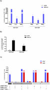
Figure 5. Longevity of the antigen specific cellular immune response induced by HIVBr18.
BALB/c mice immunized with HIVBr18 were euthanized at the indicated time points after the last immunization. Splenocytes were cultured in the presence of pooled HIV-1 peptides. Percentage of proliferating CD3+CD4+ (A) and CD3+CD8+ (B) T cells, 2 (white bar), four (dark gray bar), twelve (black bar) and 24 weeks (striped bar) after last injection. Values of proliferating (CFSElow) CD3+CD4+ T cells from the pVAX1 group after background subtraction were 0.11%; 0.24%; 0% and 0.03% at 2, 4, 12 and 24 weeks respectively. Values of CD3+CD8+ CFSElow cells were always below 0.1%; (C) Frequencies of IFNγ and IL-2- secreting cells as measured by ELISPOT assay. Values from the pVAX1 immunized group were always below 5 SFU/106 cells; (D) After 48 hours of incubation with pooled HIV-1 peptides, culture supernatants were analyzed for the presence of IFNγ, TNFα, IL-2, IL-4 and IL-5 using the mouse Th1/Th2 cytokine cytometric bead array (CBA). NT = not tested; *p<0.05, **p<0.01, ***p<0.001.
Figure 6
Figure 6. Memory T cell kinetics after immunization with HIVBr18.
Two and 24 weeks after the last immunization with HIVBr18 or the empty vector pVAX1, spleen cells from 6 BALB/c mice were cultured in the presence of pooled HIV-1 peptides. Simultaneous assessment of antigen-specific cytokine production and T cell memory phenotype was performed in gated CD3+CD4+ cells. Analysis of CD44 and CD62L expression was determined on gated CD3+CD4+ cells. IFNγ and/or IL-2 producing effector (CD44hi CD62Llow) and central memory (CD44hi CD62Lhi) CD4+ T cells are depicted.

Similar articles

Cited by

References

    1. Watkins DI, Burton DR, Kallas EG, Moore JP, Koff WC. Nonhuman primate models and the failure of the Merck HIV-1 vaccine in humans. Nat Med. 2008;14:617–621. - PMC - PubMed
    1. Buchbinder SP, Mehrotra DV, Duerr A, Fitzgerald DW, Mogg R, et al. Efficacy assessment of a cell-mediated immunity HIV-1 vaccine (the Step Study): a double-blind, randomised, placebo-controlled, test-of-concept trial. Lancet. 2008;372:1881–1893. - PMC - PubMed
    1. Corey L, McElrath MJ, Kublin JG. Post-step modifications for research on HIV vaccines. AIDS. 2009;23:3–8. - PMC - PubMed
    1. Sekaly RP. The failed HIV Merck vaccine study: a step back or a launching point for future vaccine development? J Exp Med. 2008;205:7–12. - PMC - PubMed
    1. McElrath MJ, De Rosa SC, Moodie Z, Dubey S, Kierstead L, et al. HIV-1 vaccine-induced immunity in the test-of-concept Step Study: a case-cohort analysis. Lancet. 2008;372:1894–1905. - PMC - PubMed

Publication types