

Interhemispheric interactions between the human primary somatosensory cortices
- PMID: 21347308
- PMCID: PMC3037378
- DOI: 10.1371/journal.pone.0016150
Interhemispheric interactions between the human primary somatosensory cortices
Abstract
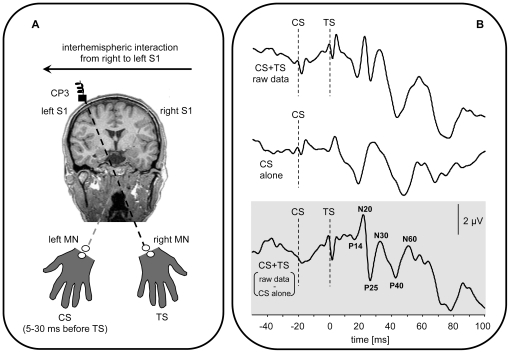
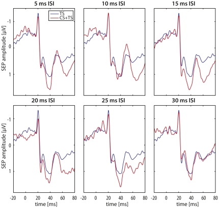
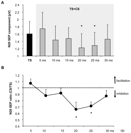
In the somatosensory domain it is still unclear at which processing stage information reaches the opposite hemispheres. Due to dense transcallosal connections, the secondary somatosensory cortex (S2) has been proposed to be the key candidate for interhemispheric information transfer. However, recent animal studies showed that the primary somatosensory cortex (S1) might as well account for interhemispheric information transfer. Using paired median nerve somatosensory evoked potential recordings in humans we tested the hypothesis that interhemispheric inhibitory interactions in the somatosensory system occur already in an early cortical processing stage such as S1. Conditioning right S1 by electrical median nerve (MN) stimulation of the left MN (CS) resulted in a significant reduction of the N20 response in the target (left) S1 relative to a test stimulus (TS) to the right MN alone when the interstimulus interval between CS and TS was between 20 and 25 ms. No such changes were observed for later cortical components such as the N20/P25, N30, P40 and N60 amplitude. Additionally, the subcortically generated P14 response in left S1 was also not affected. These results document the existence of interhemispheric inhibitory interactions between S1 in human subjects in the critical time interval of 20-25 ms after median nerve stimulation.
Conflict of interest statement
Figures
Similar articles
-
Exploring the specific time course of interhemispheric inhibition between the human primary sensory cortices.J Neurophysiol. 2014 Sep 15;112(6):1470-6. doi: 10.1152/jn.00074.2014. Epub 2014 Jun 18. J Neurophysiol. 2014. PMID: 24944212
-
Bilateral median nerve stimulation and High-Frequency Oscillations unveil interhemispheric inhibition of primary sensory cortex.Clin Neurophysiol. 2024 Sep;165:154-165. doi: 10.1016/j.clinph.2024.06.011. Epub 2024 Jun 29. Clin Neurophysiol. 2024. PMID: 39033697
-
Cortical contributions to sensory gating in the ipsilateral somatosensory cortex during voluntary activity.J Physiol. 2017 Sep 15;595(18):6203-6217. doi: 10.1113/JP274504. Epub 2017 Aug 18. J Physiol. 2017. PMID: 28513860 Free PMC article.
-
Potentials evoked in human and monkey cerebral cortex by stimulation of the median nerve. A review of scalp and intracranial recordings.Brain. 1991 Dec;114 ( Pt 6):2465-503. doi: 10.1093/brain/114.6.2465. Brain. 1991. PMID: 1782527 Review.
-
Linking 600-Hz "spikelike" EEG/MEG wavelets ("sigma-bursts") to cellular substrates: concepts and caveats.J Clin Neurophysiol. 2000 Jul;17(4):377-96. doi: 10.1097/00004691-200007000-00004. J Clin Neurophysiol. 2000. PMID: 11012041 Review.
Cited by
-
Two Distinct Neural Mechanisms Underlying Acupuncture Analgesia.Front Pain Res (Lausanne). 2022 May 18;3:869884. doi: 10.3389/fpain.2022.869884. eCollection 2022. Front Pain Res (Lausanne). 2022. PMID: 35663250 Free PMC article.
-
Visual responsiveness in sensorimotor cortex is increased following amputation and reduced after mirror therapy.Neuroimage Clin. 2019;23:101882. doi: 10.1016/j.nicl.2019.101882. Epub 2019 May 30. Neuroimage Clin. 2019. PMID: 31226622 Free PMC article.
-
Understanding the role of the primary somatosensory cortex: Opportunities for rehabilitation.Neuropsychologia. 2015 Dec;79(Pt B):246-55. doi: 10.1016/j.neuropsychologia.2015.07.007. Epub 2015 Jul 9. Neuropsychologia. 2015. PMID: 26164474 Free PMC article.
-
Interaction of the Left-Right Somatosensory Pathways in Patients With Thalamic Hemorrhage: A Case Report.Front Hum Neurosci. 2021 Nov 1;15:761186. doi: 10.3389/fnhum.2021.761186. eCollection 2021. Front Hum Neurosci. 2021. PMID: 34790107 Free PMC article.
-
The Role of Interhemispheric Interactions in Cortical Plasticity.Front Neurosci. 2021 Jul 9;15:631328. doi: 10.3389/fnins.2021.631328. eCollection 2021. Front Neurosci. 2021. PMID: 34305511 Free PMC article.
References
-
- Chen R. Interactions between inhibitory and excitatory circuits in the human motor cortex. Exp Brain Res. 2004;154:1–10. - PubMed
-
- Voller B, Floel A, Werhahn KJ, Ravindran S, Wu CW, et al. Contralateral hand anesthesia transiently improves poststroke sensory deficits. Ann Neurol. 2006;59:385–388. - PubMed
-
- Hoechstetter K, Rupp A, Stancak A, Meinck HM, Stippich C, et al. Interaction of tactile input in the human primary and secondary somatosensory cortex–a magnetoencephalographic study. Neuroimage. 2001;14:759–767. - PubMed
-
- Stancak A, Hoechstetter K, Tintera J, Vrana J, Rachmanova R, et al. Source activity in the human secondary somatosensory cortex depends on the size of corpus callosum. Brain Res. 2002;936:47–57. - PubMed
MeSH terms
LinkOut - more resources
Full Text Sources
Research Materials
