. 2011 Feb 10;7(2):e1001275.
doi: 10.1371/journal.ppat.1001275.

Epstein-Barr virus nuclear antigen 3C facilitates G1-S transition by stabilizing and enhancing the function of cyclin D1

Affiliations

Epstein-Barr virus nuclear antigen 3C facilitates G1-S transition by stabilizing and enhancing the function of cyclin D1

Abhik Saha et al. PLoS Pathog. .

Abstract

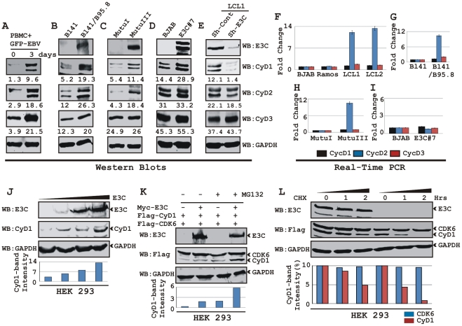
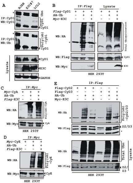
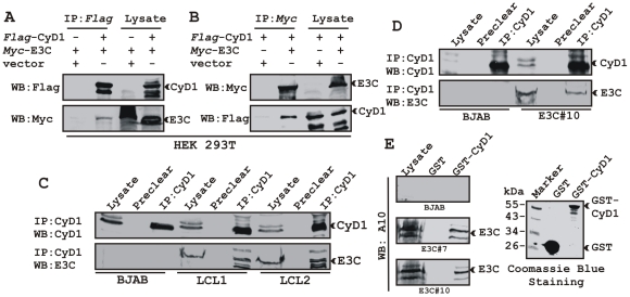
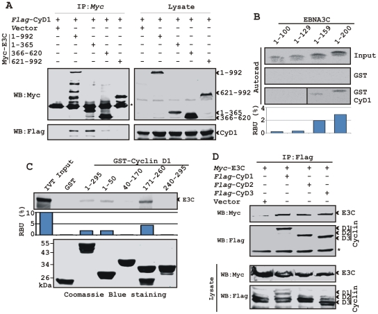
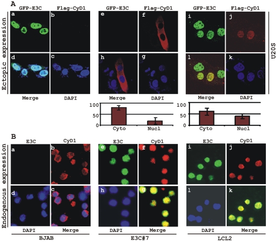
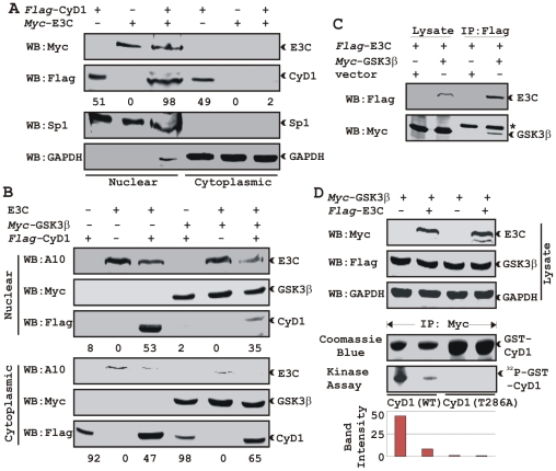
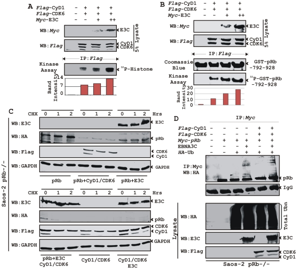
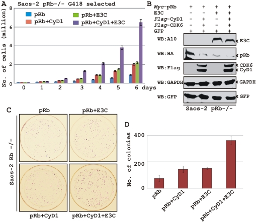
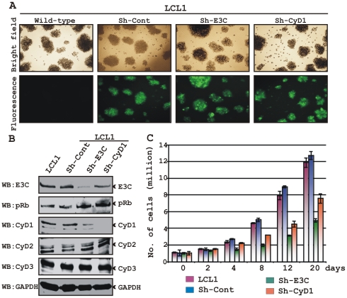
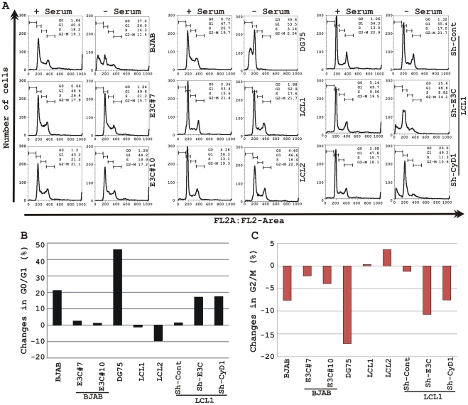
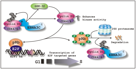
EBNA3C, one of the Epstein-Barr virus (EBV)-encoded latent antigens, is essential for primary B-cell transformation. Cyclin D1, a key regulator of G1 to S phase progression, is tightly associated and aberrantly expressed in numerous human cancers. Previously, EBNA3C was shown to bind to Cyclin D1 in vitro along with Cyclin A and Cyclin E. In the present study, we provide evidence which demonstrates that EBNA3C forms a complex with Cyclin D1 in human cells. Detailed mapping experiments show that a small N-terminal region which lies between amino acids 130-160 of EBNA3C binds to two different sites of Cyclin D1- the N-terminal pRb binding domain (residues 1-50), and C-terminal domain (residues 171-240), known to regulate Cyclin D1 stability. Cyclin D1 is short-lived and ubiquitin-mediated proteasomal degradation has been targeted as a means of therapeutic intervention. Here, we show that EBNA3C stabilizes Cyclin D1 through inhibition of its poly-ubiquitination, and also increases its nuclear localization by blocking GSK3β activity. We further show that EBNA3C enhances the kinase activity of Cyclin D1/CDK6 which enables subsequent ubiquitination and degradation of pRb. EBNA3C together with Cyclin D1-CDK6 complex also efficiently nullifies the inhibitory effect of pRb on cell growth. Moreover, an sh-RNA based strategy for knock-down of both cyclin D1 and EBNA3C genes in EBV transformed lymphoblastoid cell lines (LCLs) shows a significant reduction in cell-growth. Based on these results, we propose that EBNA3C can stabilize as well as enhance the functional activity of Cyclin D1 thereby facilitating the G1-S transition in EBV transformed lymphoblastoid cell lines.


Conflict of interest statement

The authors have declared that no competing interests exist.

Figures

Figure 1
Figure 1. EBV nuclear antigen EBNA3C stabilizes Cyclin D1 protein level.
A) 10 million human peripheral blood mononuclear cells (PBMC) were infected by BAC GFP-EBV for 4 h. At 3-days post-infection cells were lysed in RIPA buffer and western blots of endogenous proteins were probed with the indicated antibodies. B-E) 20 million cells of (B) Burkitt's lymphoma (BL) cell line BL41 and BL41 cells infected with wild-type EBV strain B95.8 (BL41/B95.8); (C) type I and III latency BL cell lines - MutuI cells (latency I gene expression program) and MutuIII cells (latency III gene expression program); (D) BJAB cells and BJAB cells stably expressing EBNA3C (BJAB_E3C#7); (E) EBNA3C and cyclin D1 knock-down LCL1 cells (LCL1_Sh-E3C and LCL1_Sh-CyD1 respectively) were harvested and total cell proteins were subjected to Western blot (WB) analysis using indicated antibodies. F-I) Total RNA was isolated from cells F) BJAB, Ramos, LCL1 and LCL2; G) BL41 and BL41/B95.8; H) MutuI and MutuIII; I) BJAB and BJAB E3C3 7 and were individually subjected to quantitative real-time PCR analysis for detecting cyclin D1, D2 and D3 transcript levels. Each sample was tested in triplicate and data obtained from three independent experiments were expressed as the difference of the quantity of specific transcripts to the quantity of GAPDH transcript as control. The fold change in expression of each cyclin D mRNA relative to GAPDH was calculated based on the threshold cycle (Ct) as 2- Δ(ΔCt), where ΔCt  =  Cttarget – CtGAPDH and Δ(ΔCt)  =  ΔCttest sample - ΔCtcontrol sample. J) HEK 293 cells were transfected with an increasing amount (0, 2, 5, 20 µg) of EBNA3C expressing construct and western blot analysis was performed to detect EBNA3C, Cyclin D1 and GAPDH. K) HEK 293 cells were co-transfected with flag-Cyclin D1 and either vector control (lanes 1 and 3) or myc-EBNA3C (lanes 2 and 4). At 36 h posttransfection, samples were treated with either 40 µM MG132 (+ lanes) or DMSO (- lanes) for 6 h and resolved by 10% SDS-PAGE and probed with the indicated antibodies. L) HEK 293 cells were similarly transfected with expression plasmids for flag-tagged both Cyclin D1 and CDK6 and myc-tagged EBNA3C as indicated. At 36 h post-transfection, cells were treated with 40 µg/ml cyclohexamide (CHX) for indicated lengths of time. 10% of the lysate from each sample were resolved by 10% SDS-PAGE. GAPDH blot was done for loading control. Western blotting was done by stripping and reprobing the same membrane. Protein bands were quantified using Odyssey imager software as indicated either as arbitrary numerical values at the bottom of gel (A-E) or as bar diagrams (J-L) based on GAPDH loading control.
Figure 2
Figure 2. EBNA3C stabilizes Cyclin D1 through inhibiting its poly-ubiquitination.
A) 50 million EBV negative BJAB cells, BJAB cells stably expressing EBNA3C (BJAB_E3C#7) and an EBV transformed cell, LCL2 were harvested after 6h incubation with proteasome inhibitor MG132 (20 µM). Cells were lysed and Cyclin D1 was immunoprecipitated (IP). Samples were resolved by 10% SDS-PAGE. Western blotting (WB) was done by stripping and reprobing the same membrane. B-E) 15 million HEK 293T cells were transiently transfected with different combinations of expression plasmids as indicated. Cells were harvested at 36h, and total protein was immunoprecipitated (IP) with indicated antibody and samples were resolved by 10% SDS-PAGE. Western blotting was done by stripping and reprobing the same membrane. Asterisks (*) indicate the immunoglobulin bands and poly-(ub) indicates poly-ubiquitination.
Figure 3
Figure 3. EBNA3C forms a complex with Cyclin D1 in human cells.
A-B) 15 million HEK 293T cells were co-transfected with myc-tagged EBNA3C and flag-tagged Cyclin D1 vectors. In each case control samples were balanced with empty vector. Cells were harvested at 36 h post-transfection and approximately 5% of the lysed cells were saved as input and the residual lysate was immunoprecipitated (IP) with 1 µg of indicated antibody. Lysates and IP complexes were resolved by 10% SDS-PAGE and western blotted (WB) with the indicated antibodies. C) 50 million BJAB cells and two different clones of EBV transformed lymphoblastoid cell lines - LCL1 and LCL2, and in D) BJAB cells stably expressing EBNA3C (BJAB_E3C#10) along with BJAB control cells were collected and lysed in RIPA buffer. Protein complexes were immunoprecipitated with Cyclin D1 specific antibody and samples were resolved by a 10% SDS-PAGE followed by western blot with antibodies as indicated. E) Either GST control or GST-cyclin D1 beads were incubated with lysates prepared from 50 million BJAB cells and two different clones of BJAB cells stably expressing EBNA3C (BJAB_E3C#7 and #10). EBNA3C was detected by western blot with the specific monoclonal antibody (A10). Coomassie staining of a 12% SDS-PAGE resolved purified GST and GST-Cyclin D1 proteins used in this study is shown in the right panel.
Figure 4
Figure 4. A small N-terminal region of EBNA3C binds to two different sites of Cyclin D1.
A) 15 million HEK 293T cells were co-transfected with vectors expressing flag-tagged Cyclin D1 and myc-tagged EBNA3C including full-length EBNA3C (residues 1-992) or different truncated mutants (residues 1–365, 366–620 and 621–992), as indicated. Cells were harvested at 36 h and 5% of the lysed cells were saved as input and the residual lysate was immunoprecipitated (IP) with 1 µg anti-myc antibody. Samples were resolved by 10% SDS-PAGE and transferred to 0.45 µm nitrocellulose membrane. The membrane was probed with flag antibody to detect co-immunoprecipitated Cyclin D1. The membrane was stripped and reprobed with anti-myc antibody to check the IP efficiency. B) Different truncated mutant constructs of EBNA3C (residues 1–100, 1–129, 1–159 and 1–200) were in vitro translated using a T7-TNT translation kit. All S35-radiolabeled in vitro translated proteins in binding buffer were precleared by rotating with GST-beads for 1 h at 4°C. Binding reactions were setup by incubating the in vitro translated proteins with either GST control or GST-Cyclin D1 overnight. Reaction samples were resolved by 15% SDS-PAGE, exposed to phosphoimager plate and scanned on a Storm 850 imaging system. C) A series of N- and C-terminal deletion mutants of GST-fused Cyclin D1 protein were purified and tested for their ability to bind in vitro translated S35-radiolabeled full-length EBNA3C as similar to (B). Coomassie staining of SDS-PAGE resolved purified GST proteins is shown in the bottom panel of (C). D) 15 million HEK 293T cells were co-transfected with myc-tagged EBNA3C and either flag-tagged Cyclin D1, Cyclin D2 or Cyclin D3. Cells were harvested at 36 h post-transfection and subjected to immunoprecipitation with 1 ug myc antibody. Lysates and IP complexes were resolved by 10% SDS-PAGE and western blotted (WB) with the indicated antibodies. Asterisks (*) indicate the immunoglobulin bands.
Figure 5
Figure 5. EBNA3C expression leads to an increase in nuclear retention of Cyclin D1.
A) U2OS cells plated on coverslips and transiently transfected with GFP-EBNA3C and flag-Cyclin D1 using Lipofectamine 2000. B) BJAB, BJAB cells stably expressing EBNA3C (BJAB_E3C#7) and an EBV transformed lymphoblastoid cell line, LCL1 were plated and air-dried onto slides. Cells were fixed using a 1∶1 mixture of acetone and methanol. Ectopically and endogenously expressed Cyclin D1 was detected using M2-antibody (1∶200 dilution) and DCS-6 (1∶50 dilution) respectively, followed by anti-mouse Alexa Fluor 594 (red). Endogenous EBNA3C in stable cell line and in EBV positive cells was detected using an EBNA3C-reactive rabbit serum (1∶150 dilution) followed by anti-rabbit Alexa Fluor 488 (green). The nuclei were counterstained using DAPI (4′,6′-diamidino-2-phenylindole). The images were sequentially captured using an Olympus confocal microscope. In (A) the bar diagram represents the mean value of 10 different fields of three independent experiments of Cyclin D1 cytoplasmic and nuclear localization.
Figure 6
Figure 6. EBNA3C bypasses GSK3β dependent nuclear export of Cyclin D1.
A) 15 million HEK 293 cells were transiently co-transfected with vectors of flag-tagged Cyclin D1 and myc-tagged EBNA3C and subjected to sub-cellular fractionation. Fractionated proteins were analyzed by probing western blots with flag and myc antibodies. Nuclear protein Sp1 and cytoplasmic protein GAPDH were immuno-detected as control. B) As similar to (A) 15 million HEK 293 cells were transfected with Cyclin D1 and different combinations of expression constructs as indicated and subjected to sub-cellular fractionation. The fractionated proteins were analyzed by using indicated antibodies. The percent distribution of Cyclin D1 is shown as numbers below panel (A) and (B). C) 15 million HEK 293T cells were co-transfected with myc-tagged GSK-3β and flag-tagged EBNA3C expression vectors. Cells proteins were collected 36 h post-transfection and immunoprecipitated (IP) with 1 ug of flag antibody. Lysates and IP complexes were resolved by 10% SDS-PAGE and western blotted (WB) with the indicated antibodies. Asterisk indicates the immunoglobulin bands. D) HEK 293T cells were transfected with myc-tagged GSK-3β and flag-tagged EBNA3C vectors as indicated. Empty vector was used to balance total transfected DNA. At 36 h posttransfction, GSK-3β immunoprecipitates were captured with myc antibody and assayed for in vitro kinase activity toward either recombinant GST-Cyclin D1 (lanes 1 and 2) or GST-Cyclin D1 T286 mutant (lanes 3 and 4) using γP32-ATP. Western blot using whole cell lysates are shown in first three panels and coomassie staining of SDS-PAGE resolved recombinant GST proteins used in this study is shown in panel 4.
Figure 7
Figure 7. EBNA3C enhances functional activity of Cyclin D1/CDK6 complex to negatively regulate pRb protein stability.
A–B) Analysis of Cyclin D1/CDK6 mediated phosphorylation of Histone H1 and pRb. HEK 293T cells were transfected with flag-tagged Cyclin D1 and CDK6 vectors and 0, 5, or 15 µg of myc-tagged EBNA3C vector. 36 h post-transfection, flag immunoprecipitates were captured and assayed for in vitro kinase activity on (A) Histone H1 or (B) recombinant GST-pRb (residues 792-928) as similar to figure 6D. C) Stability assay of pRb. Saos-2 (pRb-/-) cells were co-transfected with expression plasmids for myc- tagged pRb, flag-tagged Cyclin D1 and CDK6, and EBNA3C. 36 h post-transfection, cells were treated with 40 µg/ml cycloheximide (CHX) for the indicated times. Samples were resolved by SDS-PAGE. GAPDH was immunodetected to normalize protein levels. Western blots were probed with the indicated antibodies. D) Ubiquitination of pRb. HEK 293T cells were transfected with expression plasmids for myc-tagged pRb, HA-tagged ubiquitin (Ub), and EBNA3C (E3C), and flag-tagged Cyclin D1/CDK6 as indicated. Cells were harvested at 36 h, and total protein was immunoprecipitated (IP) with myc-specific antibody. Samples were resolved by SDS-PAGE. Western blots were probed with the indicated antibodies.
Figure 8
Figure 8. EBNA3C coupled with Cyclin D1/CDK6 complex nullifies the growth suppressive effect by pRb.
A-B) Saos-2 cells were transfected with expression plasmids for myc-tagged pRb, flag-tagged Cyclin D1 and CDK6 and EBNA3C. Cells were additionally transfected with GFP expression vector. Cells were selected for 2 weeks with G418. A) Approximately 0.1×10⁶ cells from each set of samples were plated into each well of the 6-well plates and cultured for 6 days. Viable cells from each well were counted by trypan blue exclusion method daily using an automated cell counter. For (B) 5×10⁶ cells were harvested, lysed in RIPA buffer and subjected for immunoblot analyses using indicated antibodies. C–D) Saos-2 cells transfected with different combinations of expression plasmids as described in panel (A) and selected similarly as stated above with G418. After a 2-week selection, cells were fixed on the plates with 4% formaldehyde and stained with 0.1% crystal violet. The area of stained cells in each dish was calculated by Image J software. A and D) The bar diagram represents the average data of two independent experiments with standard deviation.
Figure 9
Figure 9. Both EBNA3C and Cyclin D1 are required for cell-cycle progression in EBV transformed cells.
(A) Lentivirus transduced short hairpin RNA vectors knock down EBNA3C and Cyclin D1 in EBV transformed LCLs. Transduction with sh-RNA-containing lentivirus and selection of EBV-infected cells (LCL1) with puromycin resulted in stable cell lines expressing specific si-RNA against EBNA3C (LCL1_sh-E3C), cyclin D1 (LCL1_sh-CyD1) and sh-RNA sequence that lacks any complementary sequences in the human genome (LCL1_sh-Cont). The selected cells with GFP fluorescence were monitored by fluorescent microscopy. (B) Western blots showing the expression levels of EBNA3C, pRb, Cyclin D1, Cyclin D2 and Cyclin D3 in LCLs. GAPDH was used as the loading control. (C) Approximately 1 million cells were plated into each well of the 6-well plates and cultured at 37°C in complete medium without puromycin. Viable cells from each well were counted by trypan blue exclusion method daily for twenty days using an automated cell counter. The results shown are representative of two independent experiments. Error bars show standard deviations.
Figure 10
Figure 10. EBNA3C and Cyclin D1 are critical for G1 to S phase progression.
A) Cells were grown for 12 h in RPMI medium containing 10% FBS (+ serum) or 0.1% FBS (- serum). Propidium iodide stained cells were analyzed by flow cytometry. The bar diagram represents the change in cell-cycle profile either in (B) G0-G1 phase or (C) G2-M phase due to serum starvation of cells. The results shown are representative of two independent experiments.
Figure 11
Figure 11. A schematic illustration of how EBNA3C regulates Cyclin D1 stability and functions to facilitate G1 to S phase transition in EBV positive cells.
In EBV transformed cells, EBNA3C forms a complex with Cyclin D1 and augments its stability through inhibiting poly-ubiquitination and blocking GSK3β activity. EBNA3C further enhances the kinase activity of Cyclin D1/CDK6 complex and recruits its activity to facilitate the ubiquitination and subsequent degradation of hyperphosphorylated form of pRb, which in turn releases E2F transcription factor from an inhibitory constraint and enables the expression of genes required for entry into the S phase.

Similar articles

Cited by

References

    1. Robertson ES. Norwich, United Kingdom: Caister Academic Press; 2010. Epstein-Barr virus: latency and transformation.
    1. Kieff E, Rickinson AB. Epstein-Barr virus and its replication. In: Knipe D, Howley P, editors. Fields virology, 4th ed. Philadelphia: Lippincott Williams & Wilkins; 2002. pp. 2511–2573.
    1. O'Nions J, Allday MJ. Deregulation of the cell cycle by the Epstein-Barr virus. Adv Cancer Res. 2004;92:119–186. - PubMed
    1. Bajaj BG, Murakami M, Cai Q, Verma SC, Lan K, et al. Epstein-Barr virus nuclear antigen 3C interacts with and enhances the stability of the c-Myc oncoprotein. J Virol. 2008;82:4082–4090. - PMC - PubMed
    1. Bain M, Watson RJ, Farrell PJ, Allday MJ. Epstein-Barr virus nuclear antigen 3C is a powerful repressor of transcription when tethered to DNA. J Virol. 1996;70:2481–2489. - PMC - PubMed

Publication types

MeSH terms