. 2011 Feb 10;7(2):e1001282.
doi: 10.1371/journal.ppat.1001282.

The C-terminus of Toxoplasma RON2 provides the crucial link between AMA1 and the host-associated invasion complex

Affiliations

The C-terminus of Toxoplasma RON2 provides the crucial link between AMA1 and the host-associated invasion complex

Jessica S Tyler et al. PLoS Pathog. .

Abstract

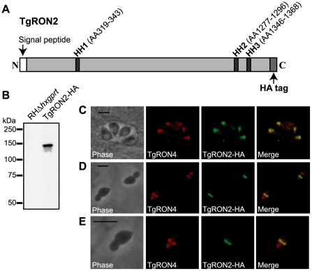
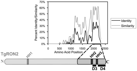
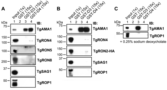
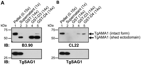
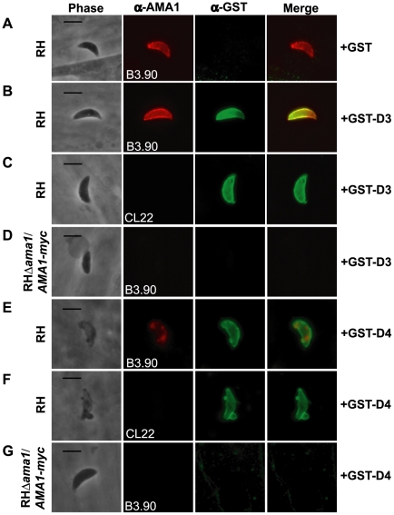
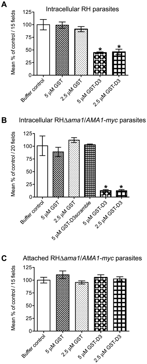
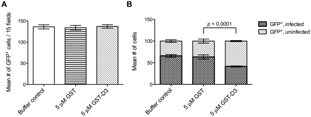
Host cell invasion by apicomplexan parasites requires formation of the moving junction (MJ), a ring-like apposition between the parasite and host plasma membranes that the parasite migrates through during entry. The Toxoplasma MJ is a secreted complex including TgAMA1, a transmembrane protein on the parasite surface, and a complex of rhoptry neck proteins (TgRON2/4/5/8) described as host cell-associated. How these proteins connect the parasite and host cell has not previously been described. Here we show that TgRON2 localizes to the MJ and that two short segments flanking a hydrophobic stretch near its C-terminus (D3 and D4) independently associate with the ectodomain of TgAMA1. Pre-incubation of parasites with D3 (fused to glutathione S-transferase) dramatically reduces invasion but does not prevent injection of rhoptry bulb proteins. Hence, the entire C-terminal region of TgRON2 forms the crucial bridge between TgAMA1 and the rest of the MJ complex but this association is not required for rhoptry protein injection.


Conflict of interest statement

The authors have declared that no competing interests exist.

Figures

Figure 1
Figure 1. Visualization of TgRON2-HA in the rhoptry necks and at the MJ.
(A) Schematic representation of the TgRON2 protein with the three putative hydrophobic helices (HH) designated by dark grey bars, with the spanning amino acids (AA) indicated above (AA positions are according to the TgRON2 sequence for the RH strain of T. gondii; Genbank accession number HQ110093). Parasites were engineered to endogenously express a derivative of TgRON2 fused to a C-terminal HA tag (TgRON2-HA). (B) Western blot analysis of the TgRON2-HA and parental parasites using the HA-specific rat monoclonal 3F10. (C, D, E) IFA analysis of infected HFF monolayers with intracellular (C) or partially-invaded (D, E) TgRON2-HA-expressing parasites. Infected monolayers were formaldehyde-fixed and permeabilized with methanol (C) or triton X-100 (D, E), then stained with rat anti-HA monoclonal 3F10 and rabbit anti-TgRON4 polyclonal sera . The images shown in (E) are enlarged views of the parasite in the bottom, left side of (D). The scale bars represent 5 µm.
Figure 2
Figure 2. Alignment of TgRON2 with its orthologues reveals greatest sequence conservation in carboxy-terminal third of protein.
The Toxoplasma RON2 polypeptide sequence was aligned with its orthologues in Neospora caninum and Plasmodium spp. (P. falciparum strain 3D7, P. berghei, P. knowlesi, and P. vivax) using ClustalX. The percent identity or percent similarity for 50 amino acid windows was calculated in 5 amino acid steps over the length of the alignment. The length of the alignment corresponds to the longest orthologue. Shown below is the relative region of TgRON2 that corresponds to the most conserved region of RON2 across the species. The regions between AA1293–1346 and AA1366–1479 were designated domain 3 (D3) and domain 4 (D4), respectively.
Figure 3
Figure 3. TgRON2 fusions GST-D3 and GST-D4 independently and specifically interact with TgAMA1 from parasite lysates.
(A) Immunoblotting analysis of NP-40-solublized RHΔhxgprt parasite lysates (Input) or material co-precipitated with molar equivalents of GST (lane 2), GST-D3 (lane 3) or GST-D4 (lane 4) was conducted to determine interaction with MJ complex members TgAMA1, TgRON4, TgRON5, and TgRON8. Western blots with each of the relevant antibodies (IB) are shown. TgSAG1 and TgROP1 serve as negative controls. Parentheses indicate loaded parasite equivalents. Size markers are indicated in kDa. (B) GST pull-down experiments were repeated as described in (A) using the TgRON2-HA parasites. (C) GST pull-down experiments were repeated as described in (A) but with supplementation of buffers with the ionic detergent sodium deoxycholate at a final concentration of 0.25% (w/v).
Figure 4
Figure 4. TgRON2 fusions GST-D3 and GST-D4 independently and specifically interact with the shed ectodomain of TgAMA1.
Extracellular parasites were incubated under conditions to induce shedding of secreted TgAMA1 into culture supernatants. Immunoblotting analysis of pelleted parasites (lane 1), cleared culture supernatants (lane 2), or material from supernatants co-precipitated with molar equivalents of GST (lane 3), GST-D3 (lane 4) or GST-D4 (lane 5) was conducted using the monoclonal antibodies B3.90 (A), specific for the TgAMA1 N-terminus, or CL22 (B), specific for the TgAMA1 C-terminus. The former detects the intact, integral membrane form (∼65 kDa) and the shed ectodomain (∼53 kDa) while CL22 detects only the intact form. TgSAG1 serves as a negative control. Parentheses indicate loaded parasite equivalents. Size markers are indicated in kDa.
Figure 5
Figure 5. TgRON2 fusions GST-D3 and GST-D4 bind to TgAMA1 on the surface of parasites.
Extracellular RH (A, B, C, E, and F) or RHΔama1/AMA1-myc (D and G) parasites were pre-treated with, and then permitted to infect HFF monolayers in the presence of, molar equivalents of GST alone (A), GST-D3 (B–D), or GST-D4 (E–G). Infected monolayers were formaldehyde-fixed and stained in the absence of permeabilization using the TgAMA1 monoclonal B3.90 (A, B, D, E, and G, panel 2), specific for the ectodomain, or the TgAMA1 monoclonal CL22 (C and F, panel 2), specific for an epitope inside the parasite cytosol, or rabbit antisera specific for GST (A–G, panel 3). Merged images for the anti-TgAMA1 and anti-GST antibodies are shown in panel 4 for each set. The RHΔama1/AMA1-myc parasites were grown under conditions known to deplete AMA1-myc expression . Scale bars represent 5 µm.
Figure 6
Figure 6. Pre-incubation of RH and RHΔama1/AMA1-myc parasites with GST-D3 decreases invasion efficiency.
(A, B) Extracellular RH (A) or RHΔama1/AMA1-myc (B) parasites were pre-treated with a buffer control or indicated molar equivalents of GST alone, GST-D3, or GST-D3scramble (B only) and then permitted to infect HFF monolayers for 15 minutes using temperature-based synchronized invasion conditions. The number of intracellular parasites was determined by differential staining of the extracellular vs. total parasites before and after detergent permeabilization. The number of intracellular parasites was determined for 15 (A) or 20 (B) randomly-selected fields from three coverslips for each condition tested. The invasion levels for each condition are shown relative to the buffer-treated control (shown are means with standard deviation). An asterisk indicates a statistically significant reduction in invasion relative to the GST controls (unpaired Student's t-test), with p<0.0099 (A) or p<0.0002 (B). (C) To determine if GST-D3 treatment affects attachment, RHΔama1/AMA1-myc parasites were pre-treated with molar equivalents of GST alone, GST-D3, or a buffer control as described above and then permitted to attach to formaldehyde-fixed HFF monolayers. The number of stained, attached parasites was counted in 15 randomly-selected fields from three coverslips for each condition tested. The attachment levels for each condition are shown relative to the buffer-treated control (shown are means with standard deviation).
Figure 7
Figure 7. Pre-incubation of parasites with GST-D3 does not affect rhoptry bulb secretion.
Extracellular SeCreEt parasites were analyzed for their ability to inject the rhoptry fusion protein toxofilin-Cre into Cre-reporter host cells following treatment with a buffer control, GST, or GST-D3. (A) The number of GFP-positive cells was determined in 15 randomly selected fields from four coverslips for each condition tested (shown are means with standard deviation). (B) The ratio of uninfected to infected GFP-positive cells, counting a total of 100 cells, was determined in randomly selected fields from four coverslips for each condition tested (shown are means with standard deviation).

References

    1. Carruthers VB, Sibley LD. Sequential protein secretion from three distinct organelles of Toxoplasma gondii accompanies invasion of human fibroblasts. Eur J Cell Biol. 1997;73:114–123. - PubMed
    1. Dobrowolski JM, Sibley LD. Toxoplasma invasion of mammalian cells is powered by the actin cytoskeleton of the parasite. Cell. 1996;84:933–939. - PubMed
    1. Keeley A, Soldati D. The glideosome: a molecular machine powering motility and host-cell invasion by Apicomplexa. Trends Cell Biol. 2004;14:528–532. - PubMed
    1. Aikawa M, Miller LH, Johnson J, Rabbege J. Erythrocyte entry by malarial parasites. A moving junction between erythrocyte and parasite. J Cell Biol. 1978;77:72–82. - PMC - PubMed
    1. Michel R, Schupp K, Raether W, Bierther FW. Formation of a close junction during invasion of erythrocytes by Toxoplasma gondii in vitro. Int J Parasitol. 1980;10:309–313. - PubMed

Publication types

MeSH terms

Substances