. 2011 Feb 9;6(2):e16951.
doi: 10.1371/journal.pone.0016951.

Rac1 targeting suppresses human non-small cell lung adenocarcinoma cancer stem cell activity

Affiliations

Rac1 targeting suppresses human non-small cell lung adenocarcinoma cancer stem cell activity

Shailaja Akunuru et al. PLoS One. .

Abstract

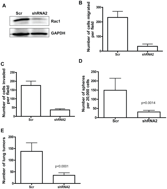
The cancer stem cell (CSC) theory predicts that a small fraction of cancer cells possess unique self-renewal activity and mediate tumor initiation and propagation. However, the molecular mechanisms involved in CSC regulation remains unclear, impinging on effective targeting of CSCs in cancer therapy. Here we have investigated the hypothesis that Rac1, a Rho GTPase implicated in cancer cell proliferation and invasion, is critical for tumor initiation and metastasis of human non-small cell lung adenocarcinoma (NSCLA). Rac1 knockdown by shRNA suppressed the tumorigenic activities of human NSCLA cell lines and primary patient NSCLA specimens, including effects on invasion, proliferation, anchorage-independent growth, sphere formation and lung colonization. Isolated side population (SP) cells representing putative CSCs from human NSCLA cells contained elevated levels of Rac1-GTP, enhanced in vitro migration, invasion, increased in vivo tumor initiating and lung colonizing activities in xenografted mice. However, CSC activity was also detected within the non-SP population, suggesting the importance of therapeutic targeting of all cells within a tumor. Further, pharmacological or shRNA targeting of Rac1 inhibited the tumorigenic activities of both SP and non-SP NSCLA cells. These studies indicate that Rac1 represents a useful target in NSCLA, and its blockade may have therapeutic value in suppressing CSC proliferation and metastasis.


Conflict of interest statement

Competing Interests: The authors have declared that no competing interests exist.

Figures

Figure 1
Figure 1. Knocking down Rac1 expression effectively inhibits human non-small cell lung cancer cell proliferation.
(A) Cell lysates collected from either scrambled shRNA (Scr) or Rac1 shRNA (shRNA1, shRNA2) A549 cells were subjected to Rac1 western blot analysis. GAPDH was used as loading control. (B) Infected cells were sorted and plated on 96-well plate and proliferation assay was performed using MTS reagent. Assay was performed in triplicates and above is one representative of three independent experiments. (C) Infected cells were sorted, plated for soft agar colony assay and colonies per field were counted after 2 weeks. Assay was performed in triplicates and above is one representative of three independent experiments. (D) Infected cells were sorted and incubated with BrdU in log phase of cell growth. Cells are trypisinized, stained with BrdU antibody, 7AAD and cell cycle analysis was performed using flowcytometry. Assay was performed in triplicates and above is one representative of four independent experiments. Error bars represents SD. (E) Control cells or cells expressing shRNA resistant Rac1 mutant were infected with scr or Rac1 shRNAs, sorted and plated on 96-well plate. Proliferation assay was performed using MTS reagent. Assay was performed in triplicates and above is one representative of two independent experiments. (F) Infected cells were sorted, plated for soft agar colony assay and colonies were counted after 10 days. Assay was performed in triplicates and above is one representative of two independent experiments.
Figure 2
Figure 2. Targeting Rac1 blocks non-small cell lung cancer cell adhesion, migration and invasion.
(A) Infected A549 cells were sorted, plated on fibronectin coated slides and stained with either p-FAK (top panel) or vinculin (middle panel) or p-Paxillin (lower panel) and DAPI. Images were collected using fluorescent microscope at 40X magnification. Images above are representative of several images obtained from two independent experiments. (B) A549 sorted cells were plated on fibronectin coated 96-well plate for in vitro adhesion assay and cells attached to plate after 1 hour was determined using MTS reagent. Adhesion assay was performed with five replicates and the data is representative of three independent experiments. (C) Sorted cells were plated on trans-well migration plates and migration of cells toward 10% FBS was measured overnight. Assay was performed in replicates and above data was representative of three independent experiments. (D) Sorted A549 cells were plated on matrigel coated invasion plates and migration of cells toward 10% FBS and 10 µg/ml fibronectin was measured after 48 hours. Assay was performed in triplicates and the above is a representative of three independent experiments. Error bars represents SD. (E) Control or cells expressing Rac1shRNA resistant mutant were infected with Rac1 shRNA and sorted. Cells were plated on trans-well migration plates and migration of cells toward 10% FBS was measured overnight. Assay was performed in triplicates and above data was representative of two independent experiments.
Figure 3
Figure 3. Knocking down Rac1 expression suppresses lung cancer cell homing and tumor growth in the lung of recipient mice.
(A) 5×10⁵ A549 cells were injected into tail vein of NSG mice (n = 6 per condition) and lungs were dissected after 8 weeks. Lung were stained with Bouins solution and destained in 70% ethanol. Quantification of lung colonization data was shown in the right panel. Error bar represents SE. Depicted is a representative of two independent experiments. (B) Tumor cell homing assay was performed as described in methods (n = 6 per condition in each experiment). Homing index was measured as percentage of YFP positive cells detected in lung, normalized to control. Depicted is a representative of three independent experiments. (C) 5×10⁵ scr or Rac1 shRNA infected cells were injected subcutaneous into flanks of NOD/SCID mice and tumor volume was measured weekly for 7 weeks. Error bar represents SE.
Figure 4
Figure 4. Side population cells possess elevated Rac1 activity, increased migration, invasion and proliferative activities, and enriched tumor initiating activity in mouse lung.
(A) A549 cells were stained with Hoechst 33342 dye and analyzed by flowcytometry for side population (left panel). Cells were treated with 10 µM Fumitremorgen for inhibitor control (right panel). Depicted is a representative of several SP analyses. (B) Cell lysates collected from sorted SP and non-SP cells were subjected to GST-PAK pull down assay and processed for Rac1 western blot analysis to determine the Rac1 activity. Total Rac1 blot was used as a control. Depicted is a representative of three independent Rac1 activity pull-down assays. (C) Sorted A549 cells were plated for trans-well migration assay and cells migrated overnight towards 10% FBS were stained and counted. Above is a representative of three independent experiments and error bars represents SD. (D) 5×10⁴ sorted SP and non-SP cells were injected into tail vein of NSG mice (n = 4 per condition). Lungs were dissected out at the end of 12 weeks. Right panel shows quantification of lung colonization data. Error bar represents SE. Above is a representative of three independent experiments.
Figure 5
Figure 5. Targeting Rac1 effectively suppresses the adhesion, migration and invasion activities of SP cells as well as non-SP cells.
(A) Sorted cells were plated on fibronectin coated slides, fixed and subjected to immunostaining with p-FAK antibody. Cell images were collected at 40X magnification using Fluorescent microscope. Above depicted are representative of multiple images collected. (B, C, D) A549 sorted cells were either plated on fibronectin coated 96-well plate for in vitro adhesion assay (B), on trans-well plates for migration assay (C) or matrigel coated invasion plates for invasion assay (D). All assays were performed in triplicates and error bars represent SD. Depicted are representative of three independent experiments.
Figure 6
Figure 6. Rac1 knockdown inhibits the proliferation and tumor seeding activities of SP cells as well as non-SP cells in vitro and in vivo.
(A) Sorted cells were plated in 96-well plate and proliferation assay was performed using MTS reagent. Assay was performed in triplicates and error bars represents SD. Depicted is a representative of three independent experiments. (B) A549 sorted cells were incubated with BrdU and cell cycle analysis was performed by BrdU staining and flowcytometric analysis. Assay was performed in triplicates and error bars represent SD. Above is a representative of two independent experiments. (C) Sorted cell were directly plated for soft agar colony formation assay and the number of colonies formed were counted after 2–3 weeks. Assay was performed in triplicates and error bars represent SD. Above is a representative of three independent experiments. (D) 50,000 sorted cells were injected into tail vein of NSG mice and the lungs were isolated for analysis after 12 weeks. Number of lung tumors was counted under light microscope and error bar represents SE. Results are representative of three independent experiments. (E) Cells were infected with either scr or Rac1 shRNA and incubated with BrdU. Cells were stained with CD133 antibody during the BrdU staining described in methods. BrdU positive cells gated from CD133+ and CD133 cells were analyzed by FACS.
Figure 7
Figure 7. Rac1 targeting inhibits primary non-small cell lung cancer cell migration, invasion, sphere forming and lung colonization activities.
(A) Cells isolated from primary tumor sample were infected with either scr or Rac1 shRNA and sorted cells were subjected to Rac1 western blot analysis. GAPDH was used as loading control. Above depicted is a representative of multiple western blots. (B, C, D) Primary human adenocarcinoma cells were plated for either trans-well migration assay (B) or invasion assay (C) or sphere assay (D). For sphere assay, sorted cells were directed plated in sphere growth media described in methods section. Number of spheres formed after 10 days were counted using light microscope. All the assays were performed in triplicates and error bar represents SD. Results are representative of three independent experiments. (E) For lung colonization assay, 5×10⁵ sorted cells were injected into tail vein of NSG mice (n = 4 per condition) and mice were sacked after 6 weeks. Number of lung tumors was counted under dissecting microscope. Error bar represents SD.

Similar articles

Cited by

References

    1. Jemal A, Siegel R, Ward E, Murray T, Xu J, et al. Cancer statistics, 2006. CA Cancer J Clin. 2006;56:106–130. - PubMed
    1. Lapidot T, Sirard C, Vormoor J, Murdoch B, Hoang T, et al. A cell initiating human acute myeloid leukaemia after transplantation into SCID mice. Nature. 1994;367:645–648. - PubMed
    1. Singh SK, Hawkins C, Clarke ID, Squire JA, Bayani J, et al. Identification of human brain tumour initiating cells. Nature. 2004;432:396–401. - PubMed
    1. Al-Hajj M, Wicha MS, Benito-Hernandez A, Morrison SJ, Clarke MF. Prospective identification of tumorigenic breast cancer cells. Proc Natl Acad Sci U S A. 2003;100:3983–3988. - PMC - PubMed
    1. O'Brien CA, Pollett A, Gallinger S, Dick JE. A human colon cancer cell capable of initiating tumour growth in immunodeficient mice. Nature. 2007;445:106–110. - PubMed

Publication types

MeSH terms

Substances