. 2011 Feb 9;6(2):e16295.
doi: 10.1371/journal.pone.0016295.

Intestinal epithelial stem/progenitor cells are controlled by mucosal afferent nerves

Affiliations

Intestinal epithelial stem/progenitor cells are controlled by mucosal afferent nerves

Ove Lundgren et al. PLoS One. .

Abstract

Background: The maintenance of the intestinal epithelium is of great importance for the survival of the organism. A possible nervous control of epithelial cell renewal was studied in rats and mice.

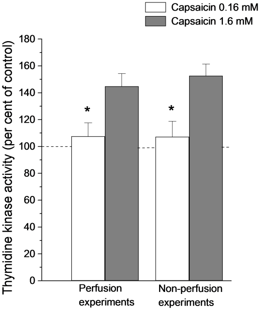
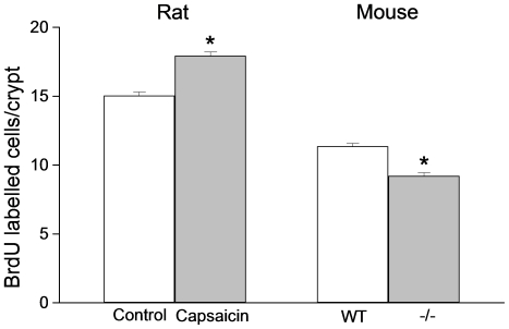
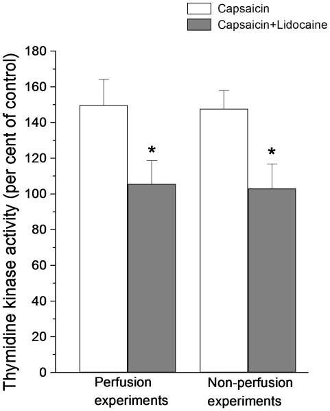
Methods: Mucosal afferent nerves were stimulated by exposing the intestinal mucosa to capsaicin (1.6 mM), which stimulates intestinal external axons. Epithelial cell renewal was investigated in the jejunum by measuring intestinal thymidine kinase (TK) activity, intestinal (3)H-thymidine incorporation into DNA, and the number of crypt cells labeled with BrdU. The influence of the external gut innervation was minimized by severing the periarterial nerves.

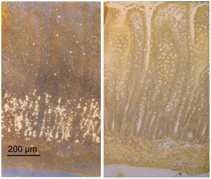
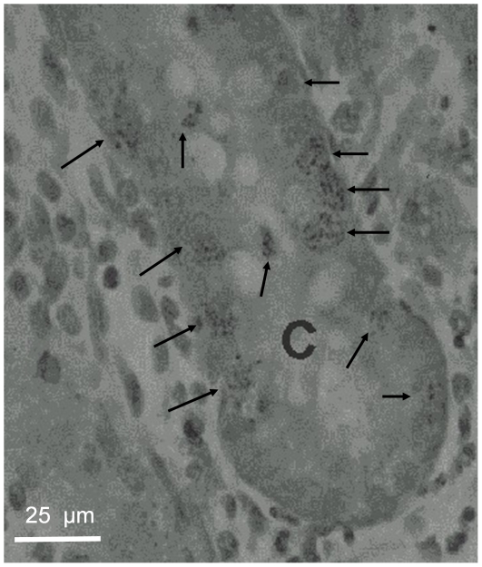
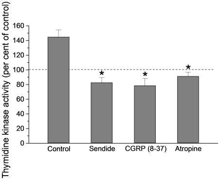
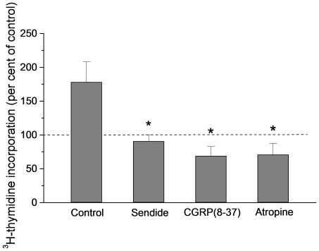
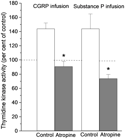
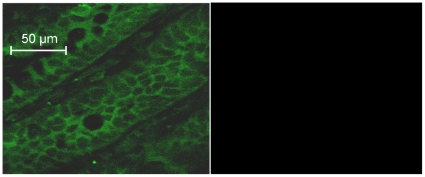
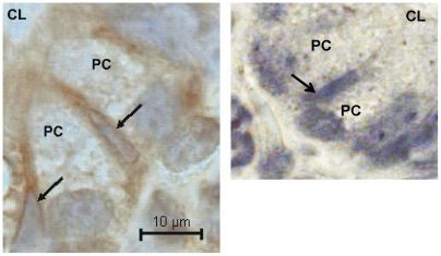
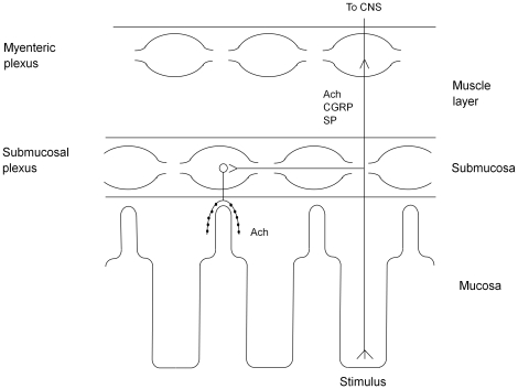
Principal findings: Luminal capsaicin increased all the studied variables, an effect nervously mediated to judge from inhibitory effects on TK activity or (3)H-thymidine incorporation into DNA by exposing the mucosa to lidocaine (a local anesthetic) or by giving four different neurotransmitter receptor antagonists i.v. (muscarinic, nicotinic, neurokinin1 (NK1) or calcitonin gene related peptide (CGRP) receptors). After degeneration of the intestinal external nerves capsaicin did not increase TK activity, suggesting the involvement of an axon reflex. Intra-arterial infusion of Substance P (SP) or CGRP increased intestinal TK activity, a response abolished by muscarinic receptor blockade. Immunohistochemistry suggested presence of M3 and M5 muscarinic receptors on the intestinal stem/progenitor cells. We propose that the stem/progenitor cells are controlled by cholinergic nerves, which, in turn, are influenced by mucosal afferent neuron(s) releasing acetylcholine and/or SP and/or CGRP. In mice lacking the capsaicin receptor, thymidine incorporation into DNA and number of crypt cells labeled with BrdU was lower than in wild type animals suggesting that nerves are important also in the absence of luminal capsaicin, a conclusion also supported by the observation that atropine lowered thymidine incorporation into DNA by 60% in control rat segments.

Conclusion: Enteric nerves are of importance in maintaining the intestinal epithelial barrier.


Conflict of interest statement

Competing Interests: The authors have declared that no competing interests exist.

Figures

Figure 1
Figure 1. Autoradiography of rat small intestine after giving tritiated thymidine i.v.
The labeled compound (50 µCi 3H-methyl-thymidine) was administred 6 h prior to removing the intestine The left panel illustrates dark field microscopy of an autoradiogram showing radiolabeled cells (white spots) almost exclusively located in the epithelial layer of the crypts of Lieberkühn. To the right the same section is seen in light microscopy. Van Gieson.
Figure 2
Figure 2. The distribution of 3H-methyl-thymidine in an intestinal crypt (C) as shown by autoradiography.
The labeled compound (50 µCi 3H-methyl-thymidine) was administred 6 h prior to removing the intestine. Only cell nuclei were found to be labeled (arrows).
Figure 3
Figure 3. BrdU labeling of the small intestine after i.v. administration.
BrdU (50 mg/kg b.wt.), a thymidine analogue, was given 2 h prior to removing the intestine. The dark labeling by BrdU is exclusively located to the nuclei of intestinal crypt cells. Mayer's haematoxylin.
Figure 4
Figure 4. The effect on intestinal thymidine kinase activity of exposing the intestinal mucosa to capsaicin (0.16 or 1.6 mM).
Thymidine kinase activity is expressed in per cent of activity measured in control segments (no capsaicin). The experiments were performed using two types of preparations. In most experiments only the proximal ends of the segments were cannulated with plastic tubing and the intestinal segment was perfused by means of a pump at a rate of 20 µL per min (“perfusion experiments”; n = 6). In other experiments the distal ends were also provided with plastic tubing closed by plugs (“non-perfusion experiments”; n = 7). Asterisks indicate statistical significance. Mean ± s.e.m.
Figure 5
Figure 5. Rat: BrdU labeled cells per crypt in intestinal segments exposed to a capsaicin or a control solution.
BrdU (50 mg/kg bwt) was given i.v. 2 h prior to removal of the intestinal segments. The number of labeled cells in 255 crypts in control (n = 5) and in 249 crypts in capsaicin segments (n = 5) were counted. Asterisk indicates statistical significance. Mean ± s.e.m. Mouse: BrdU labeled cells per crypt in wild type or capsaicin receptor knock-out mice. Five capsaicin receptor knock-out mice (−/−; 225 crypts) and in five wild type mice (WT; 215 crypts) were investigated. BrdU (100 mg/kg b.wt.) was administered i.p. 2 h before removal of the small intestine. Asterisk indicates statistical significance. Mean ± s.e.m.
Figure 6
Figure 6. The effect of capsaicin on intestinal thymidine kinase activity after anesthetizing the mucosa with lidocaine.
Thymidine kinase activity is expressed in per cent of activity measured in control segments (no capsaicin). Asterisks indicate statistical significance between the two groups of experiments (perfusion experiments, n = 8; non-perfusion experiments, n = 7). Mean ± s.e.m.
Figure 7
Figure 7. The effect of neurotransmitter receptor antagonists on capsaicin evoked increase of thymidine kinase activity.
The receptor antagonists tested were: Sendide (NK1 receptor antagonist); 8–37 α-GGRP (CGRP receptor antagonist); atropine (muscarinic receptor antagonist). Thymidine kinase activity is expressed in per cent of activity measured in control segments (no capsaicin). N = 6 for each antagonist experiments. Asterisks indicate statistical significance compared to control. Mean ± s.e.m.
Figure 8
Figure 8. The effect of neurotransmitter receptor antagonists on capsaicin evoked increase of 3H-thymidine incorporation into DNA.
The receptor antagonists tested were: Sendide (NK1 receptor antagonist); 8–37 α-GGRP (CGRP receptor antagonist); atropine (muscarinic receptor antagonist). The receptor blockers were given as described in Methods. 3H-thymidine incorporation is expressed in per cent of incorporation measured in control segments (no capsaicin). Asterisks indicate statistical significance compared to control. Mean ± s.e.m.
Figure 9
Figure 9. Thymidine kinase activity in intestinal segments after i.a. infusion of substance P or CGRP.
The peptides were infused for one hour at a rate of 10−9 mol per minute with or without prior administration of atropine (0.5 mg per kg b.wt. i.v.). Note that in these experiments thymidine kinase activity is expressed in per cent of activity measured in segments removed at time 0. Asterisks indicate statistical significance. Mean ± s.e.m.
Figure 10
Figure 10. The localization of M5 muscarinic receptor immunoreactivity in intestinal crypts.
The picture is photographed by confocal microscopy corresponding to a 4 µm thick section of the wall of the crypts. Hence, no crypt lumen is seen and the crypt cells are cross-sectioned. The left panel of the figure suggests that the M5 receptor is located to the plasma membrane of the crypt cells. Tissue sections exposed only to the secondary antibody did not exhibit any fluorescence (right panel).
Figure 11
Figure 11. The localization of M3 muscarinic receptor immunoreactivity on intestinal stem cells.
The stem cells, indicated by arrows, are slender cells located between the Paneth cells (PC) at the very base of the crypts , . The left part of the figure indicates that the muscarinic receptor M3 is located to the plasma membrane of the presumed intestinal stem cells. The tissue section in the right panel was only exposed to the secondary antibody. Mayer's haematoxylin. CL  =  crypt lumen.
Figure 12
Figure 12. Model of axon reflex control of cell renewal in the small intestine.
The model is in part based on the findings of the present study (cf. Table 1) and is consistent with known types of neurons in the intestinal wall. It implies that the direct control of the stem/progenitor cells is exerted by a cholinergic neuron, which, in turn, is under the influence of an axon reflex releasing CGRP/SP/acetylcholine. Ach: acetylcholine; CGRP: calcitonin gene related peptide; SP: substance P; CNS: central nervous system.

Similar articles

Cited by

References

    1. Moore KA, Lemischka IR. Stem cells and their niches. Science. 2006;311:1880–1885. - PubMed
    1. Furness JB. Oxford: Blackwell Publishing Ltd; 2006. The enteric nervous system.
    1. Vidrich A, Buzan JM, De La Rue SA, Cohn SA. Physiology of gastrointestinal stem cells. In Physiology of the Gastrointestinal Tract Fourth edition, In: Johnson LR, editor. Elsevier Inc., Amsterdam; 2006. pp. 307–344.
    1. Tutton PJ. The influence of cholinoceptor activity on the mitotic rate in the crypts of Lieberkűhn in rat jejunum. Clin Exp Pharmacol Physiol. 1975;2:269–76. - PubMed
    1. Tutton PJ, Barkla DH. Neural control of colonic cell proliferation. Cancer. 1980;45:1172–1177. - PubMed

Publication types

MeSH terms