. 2011 Feb 8;5(2):e955.
doi: 10.1371/journal.pntd.0000955.

A coproantigen diagnostic test for Strongyloides infection

Affiliations

A coproantigen diagnostic test for Strongyloides infection

Alex M Sykes et al. PLoS Negl Trop Dis. .

Abstract

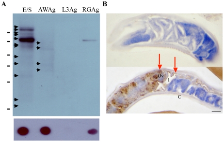
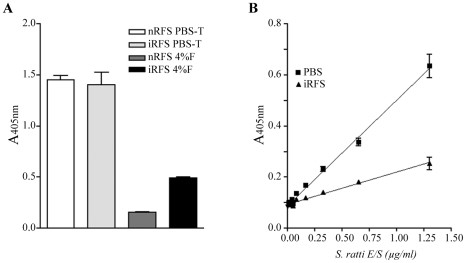
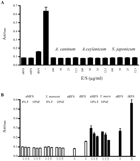
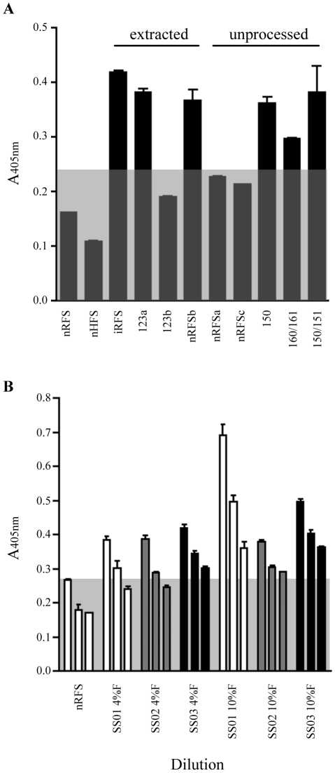
Accurate diagnosis of infection with the parasite Strongyloides stercoralis is hampered by the low concentration of larvae in stool, rendering parasitological diagnosis insensitive. Even if the more sensitive agar plate culture method is used repeated stool sampling is necessary to achieve satisfactory sensitivity. In this manuscript we describe the development of a coproantigen ELISA for diagnosis of infection. Polyclonal rabbit antiserum was raised against Strongyloides ratti excretory/secretory (E/S) antigen and utilized to develop an antigen capture ELISA. The assay enabled detection of subpatent rodent S. ratti and human S. stercoralis infection. No cross-reactivity was observed with purified E/S from Schistosoma japonicum, the hookworms Ancylostoma caninum, A. ceylanicum, nor with fecal samples collected from rodents harboring Trichuris muris or S. mansoni infection. Strongyloides coproantigens that appear stable when frozen as formalin-extracted fecal supernatants stored at -20 °C remained positive up to 270 days of storage, whereas supernatants stored at 4 °C tested negative. These results indicate that diagnosis of human strongyloidiasis by detection of coproantigen is an approach worthy of further development.


Conflict of interest statement

The authors have declared that no competing interests exist.

Figures

Figure 1
Figure 1. Immunoreactivity of the α-E/S antibody is primarily specific for adult S. ratti E/S.
α-E/S IgG recognizes primarily Strongyloides E/S antigens (A): Western blot analysis of (E/S) adult worm E/S, (AWAg) adult worm antigen, (L3Ag) 3rd stage larval antigen and (RGAg) soluble naïve rat gut antigen, using α-E/S IgG at a dilution of 5.5 µg/ml. A single immunodominant ∼75 kDa cross-reacting band was observed in the uninfected rat faecal supernatant immunoprecipitation experiment. Other immunoreactive protein bands are recognised by the α-E/S Ab and are not seen in uninfected rat gut extract. These S. ratti-specific proteins were detected at approximate molecular weights of 38 kDa, 40 kDa, 60 kDa and 70 kDa in AWAg, with additional 35 kDa, 50 kDa, 65 kDa, 100 kDa and >100 kDa bands being observed in E/S. (B) Immunoperoxidase-stained parasitic adult worm sections, probed with either (upper panel) 1∶500 preimmune rabbit serum, or (lower panel) α-ES IgG diluted 5.5 µg/ml in PBS. Scale bar  = 20 µm. Legend: I =  intestine; Ov  =  ovaries; C =  cuticle. Arrows highlight the specific ovarian and intestinal cells recognised by this antibody.
Figure 2
Figure 2. Formalin preservation allows specific discrimination of infected rat fecal supernatant from uninfected fecal supernatant.
(A) Effect of assay specificity after various treatments. Pooled uninfected fecal supernatant collected from 6 rats (nRFS) and infected rat fecal supernatant collected from 2 rats infected with S. ratti (13 and 21 dpi) (iRFS) were extracted simultaneously in PBS-T and 4% formalin (4%F). Only extraction in formalin led to a positive discrimination between nRFS and iRFS. All points are mean ± SEM of OD values obtained from duplicate samples. (B) Detection of known amounts of E/S products diluted in PBS or uninfected rat fecal supernatant (nRFS) extracted in 4% formalin. The lowest concentration of E/S products detected was 80 ng/ml and 325 ng/ml when diluted in PBS and nRFS, respectively. All points are mean ± SEM of OD values obtained from 4 samples.
Figure 3
Figure 3. No cross reactivity is observed with purified E/S antigens or faecal supernatants collected from various helminths.
(A) Testing of the Strongyloides coproELISA with E/S products collected from other helminths. All Ancylostoma and Schistosoma E/S products tested negative even at the high concentration of 100 µg/ml. Pooled uninfected rat fecal supernatant collected from 6 rats (nRFS) and infected rat fecal supernatant (iRFS) collected from two rats (13 and 21 days post infection). iRFS and 1.3 µg/ml S.ratti E/S (+) tested positive. All points are mean ± SEM of OD values obtained from 4 identical samples. (B) Specificity for Strongyloides coproantigen detection. White bars and black bars represent data from two different experiments. Diluted samples were prepared from undiluted fecal supernatant to 1∶4 by doubling dilution in PBS. Uninfected mouse fecal supernatant (nMFS), Trichuris muris and Schistosoma mansoni supernatants test negative even when applied undiluted. Uninfected rat fecal supernatant (nRFS) and infected rat fecal supernatant (iRFS) tests negative and positive, respectivly. All points are mean ± SEM of OD values obtained from duplicate samples. Assay cuttoff for negative reading is represented by the grey box.
Figure 4
Figure 4. Effect of sample storage on rat fecal supernatant and detection of heterologous S. stercoralis coproantigen.
(A) Pooled uninfected rat fecal supernatant collected from 6 rats (nRFS) and infected rat fecal supernatant (iRFS) collected from two rat (13 and 21 dpi) had previously been stored at −20°C and consistently tested negative and positive, respectively. Numbered samples 123, 150, 151, 160 and 161 represent samples collected from rats infected with known amounts of S. ratti infective larvae. Extracted fecal supernatant stored at 4°C displayed erratic absorbance values, as fecal supernatants from both uninfected and infected rats test positive. All unprocessed S. ratti infected feces stored in formalin (4% or 10%) tested positive for coproantigen when extracted after storage. Uninfected rat fecal feces tested negative when stored unextracted. All points are mean ± SEM of OD values obtained from duplicate samples. (B) Uninfected human fecal supernatant (nHFS) and Strongyloides-infected human fecal supernatants collected daily from a single patient with clinically diagnosed strongyloidiasis (SS01-SS03) were extracted simultaneously with 4% formalin and 10% formalin. All points are mean ± SEM. Assay cuttoff for negative reading is represented by the grey box.

References

    1. Dreyer G, Fernandes-Silva E, Alves S, Rocha A, Albuquerque R, et al. Patterns of detection of Strongyloides stercoralis in stool specimens: implications for diagnosis and clinical trials. J Clin Microbiol. 1996;34:2569–2571. - PMC - PubMed
    1. Sato Y, Otsuru M, Takara M, Shiroma Y. Intradermal reactions in strongyloidiasis. Int J Parasitol. 1986;16:87–91. - PubMed
    1. Page WA, Dempsey K, McCarthy JS. Utility of serological follow-up of chronic strongyloidiasis after anthelminthic chemotherapy. Trans R Soc Trop Med Hyg. 2006;100:1056–1062. - PubMed
    1. Conway DJ, Atkins NS, Lillywhite JE, Bailey JW, Robinson RD, et al. Immunodiagnosis of Strongyloides stercoralis infection: a method for increasing the specificity of the indirect ELISA. Trans R Soc Trop Med Hyg. 1993;87:173–176. - PubMed
    1. Allan JC, Craig PS. Coproantigens in gut tapeworm infections: Hymenolepis diminuta in rats. Parasitol Res. 1989;76:68–73. - PubMed

Publication types