

Controlled release of IGF-1 and HGF from a biodegradable polyurethane scaffold
- PMID: 21347565
- PMCID: PMC3200230
- DOI: 10.1007/s11095-011-0391-z
Controlled release of IGF-1 and HGF from a biodegradable polyurethane scaffold
Abstract
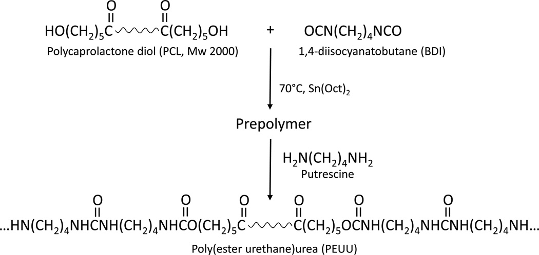
Purpose: Biodegradable elastomers, which can possess favorable mechanical properties and degradation rates for soft tissue engineering applications, are more recently being explored as depots for biomolecule delivery. The objective of this study was to synthesize and process biodegradable, elastomeric poly(ester urethane)urea (PEUU) scaffolds and to characterize their ability to incorporate and release bioactive insulin-like growth factor-1 (IGF-1) and hepatocyte growth factor (HGF).
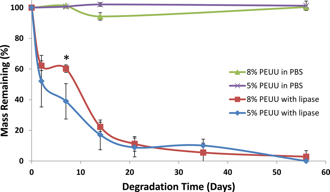
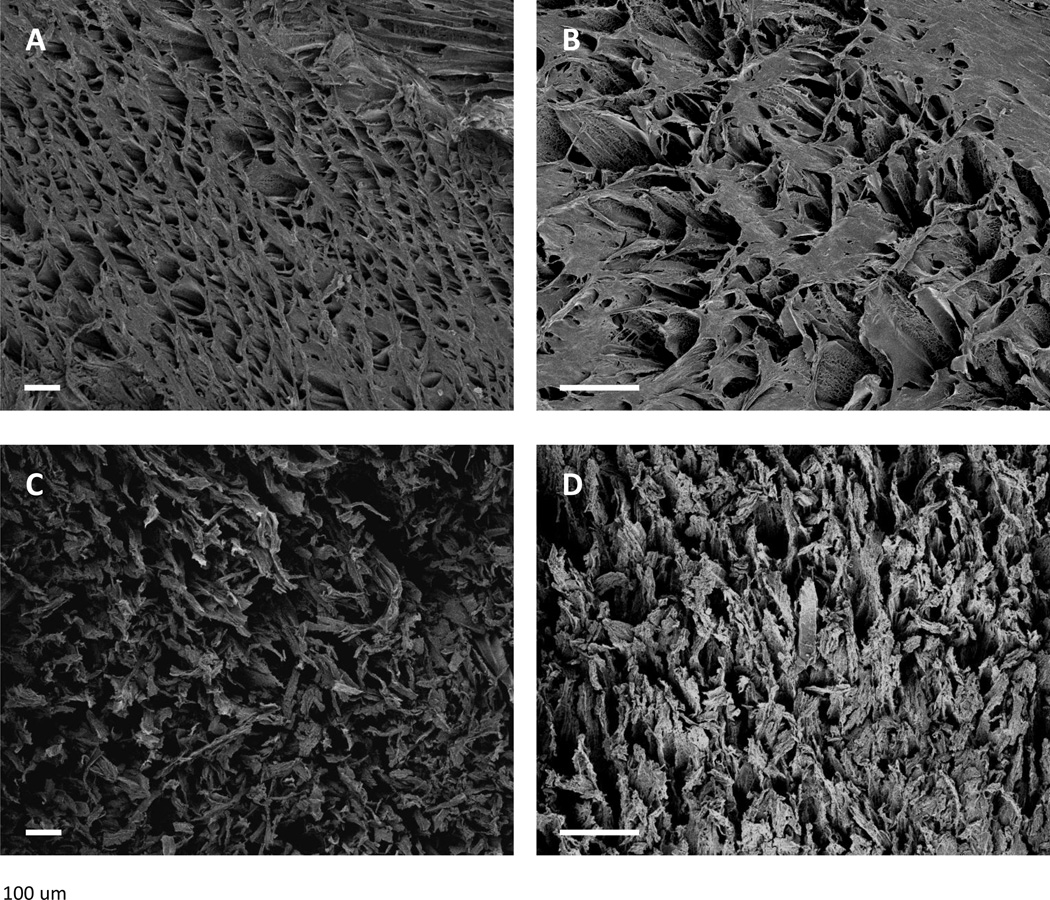
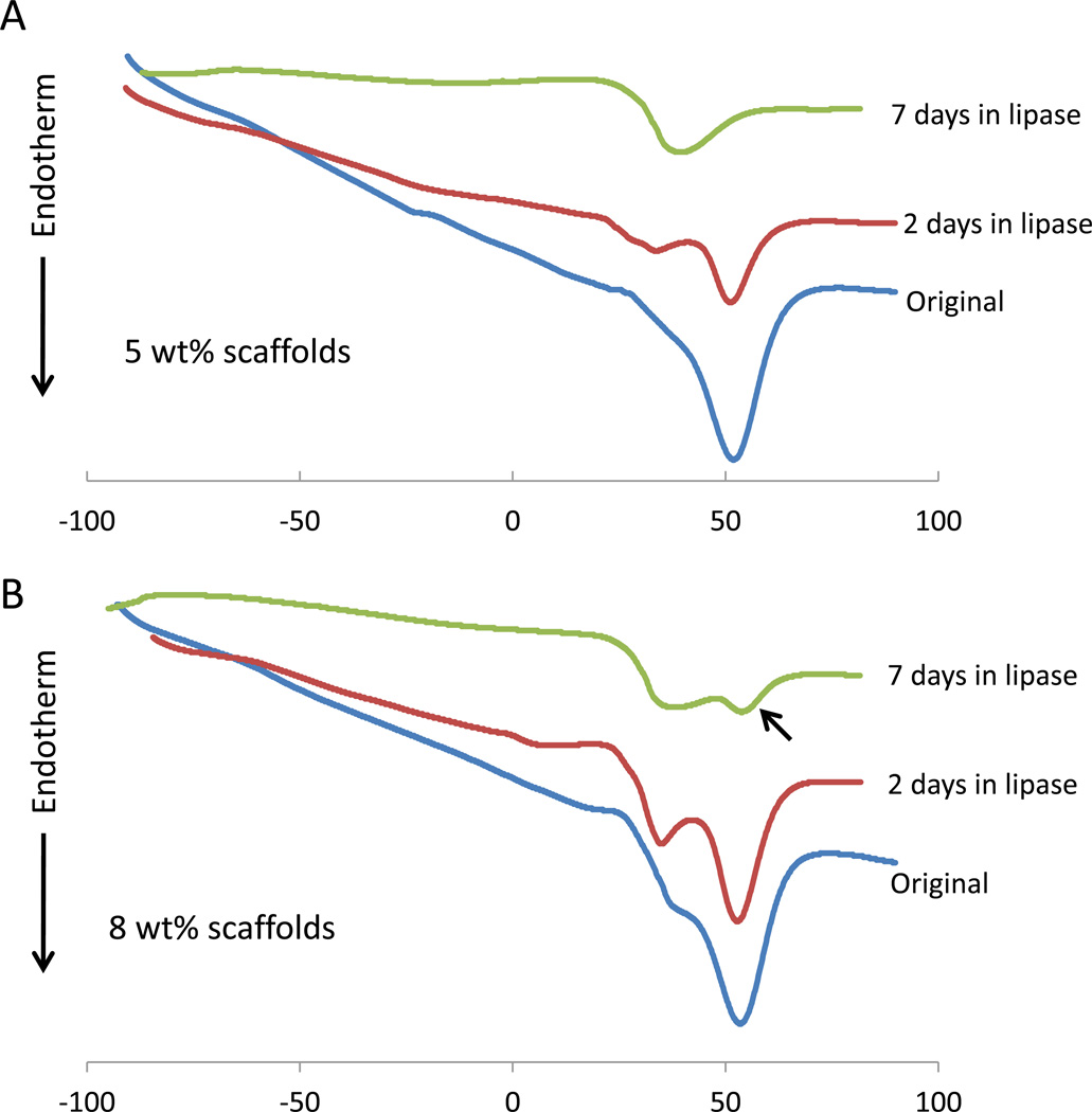
Methods: Porous PEUU scaffolds made from either 5 or 8 wt% PEUU were prepared with direct growth-factor incorporation. Long-term in vitro IGF-1 release kinetics were investigated in saline or saline with 100 units/ml lipase to simulate in vivo degradation. Cellular assays were used to confirm released IGF-1 and HGF bioactivity.
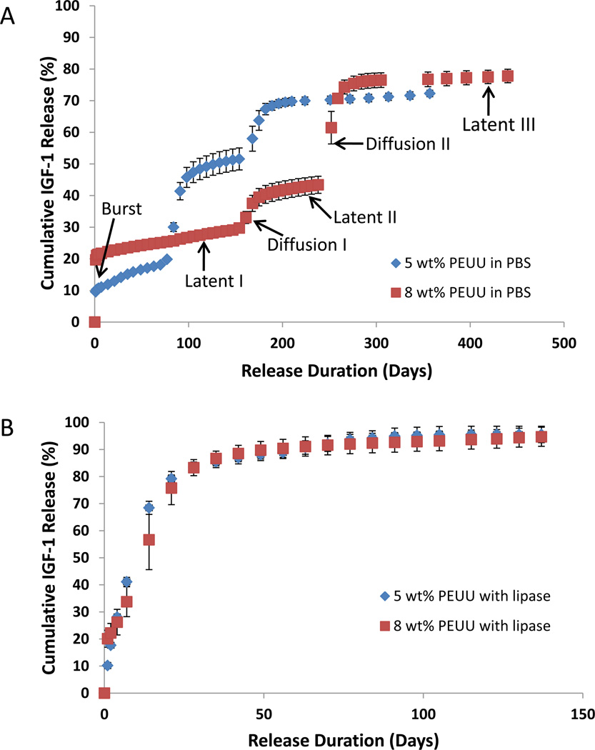
Results: IGF-1 release into saline occurred in a complex multi-phasic manner for up to 440 days. Scaffolds generated from 5 wt% PEUU delivered protein faster than 8 wt% scaffolds. Lipase-accelerated scaffold degradation led to delivery of >90% protein over 9 weeks for both polymer concentrations. IGF-1 and HGF bioactivity in the first 3 weeks was confirmed.
Conclusions: The capacity of a biodegradable elastomeric scaffold to provide long-term growth-factor delivery was demonstrated. Such a system might provide functional benefit in cardiovascular and other soft tissue engineering applications.
Figures
References
-
- Wells RG. The role of matrix stiffness in regulating cell behavior. Hepatology. 2008;47:1394–1400. - PubMed
-
- Ramaswami P, Wagner WR. Cardiovascular Tissue Engineering. In: Guelcher S, Hollinger JO, editors. An Introduction to Biomaterials. Boca Raton, FL: CRC Press; 2006. pp. 461–484.
-
- Guan J, Sacks MS, Beckman EJ, Wagner WR. Synthesis, characterization, and cytocompatibility of elastomeric, biodegradable poly(ester-urethane)ureas based on poly(caprolactone) and putrescine. J Biomed Mater Res. 2002;61:493–503. - PubMed
-
- Skarja GA, Woodhouse KA. In vitro degradation and erosion of degradable, segmented polyurethanes containing an amino acid-based chain extender. J Biomater Sci Polym Ed. 2001;12:851–873. - PubMed
Publication types
MeSH terms
Substances
Grants and funding
LinkOut - more resources
Full Text Sources
Miscellaneous
