Review
. 2011 Mar;240(1):72-91.
doi: 10.1111/j.1600-065X.2010.00990.x.

Invasion and intracellular survival by protozoan parasites

Affiliations
Review

Invasion and intracellular survival by protozoan parasites

L David Sibley. Immunol Rev. 2011 Mar.

Abstract

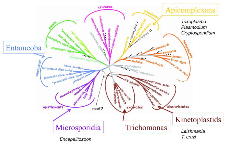
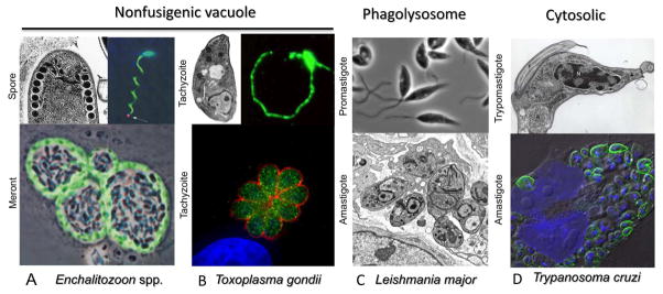
Intracellular parasitism has arisen only a few times during the long ancestry of protozoan parasites including in diverse groups such as microsporidians, kinetoplastids, and apicomplexans. Strategies used to gain entry differ widely from injection (e.g. microsporidians), active penetration of the host cell (e.g. Toxoplasma), recruitment of lysosomes to a plasma membrane wound (e.g. Trypanosoma cruzi), to host cell-mediated phagocytosis (e.g. Leishmania). The resulting range of intracellular niches is equally diverse ranging from cytosolic (e.g. T. cruzi) to residing within a non-fusigenic vacuole (e.g. Toxoplasma, Encephalitozoon) or a modified phagolysosome (e.g. Leishmania). These lifestyle choices influence access to nutrients, interaction with host cell signaling pathways, and detection by pathogen recognition systems. As such, intracellular life requires a repertoire of adaptations to assure entry-exit from the cell, as well as to thwart innate immune mechanisms and prevent clearance. Elucidating these pathways at the cellular and molecular level may identify key steps that can be targeted to reduce parasite survival or augment immunologic responses and thereby prevent disease.


Figures

Figure 1
Figure 1
Phylogenetic relationships among the eight major groups of eukaryotes. Tree is based on consensus of molecular and morphological data. Protozoan parasite groups that commonly infect humans are denoted adjacent to the major branches they occupy. Modified with permission from (1).
Figure 2
Figure 2
Summary of invasive forms and intracellular niches of major groups of intracellular protozoan parasites. A) Extracellular spore stages of E. intestinalis (left) showing coiled polar tube in cross-section. Image used with permission (216). Image at right shows discharge of sporoplasm (red) at the tip of polar tube (arrow), polar tube and spore wall stained with polyclonal serum (green), nuclei stained with DAPI (blue). Used with permission (28). Lower image shows intracellular meronts dividing within fibroblasts. Stained with mAb 6G2 that recognizes peripheral meronts (green), while sporoblasts occupy central region of the vacuole. Used with permission (28). B) Extracellular tachyzoites of T. gondii display polarized arrangement of organelles characteristic of apicomplexans (left upper). Image provided by W. Beatty. Gliding motility of tachyzoites in vitro as revealed by staining with the surface protein SGA1 (right image). Lower image shows intracellular replication of tachyzoites in a rosette. Cytoplasmic actin-like protein ALP1 (green) and surface antigen SAG1 (red) stained with specific antibodies, nuclei stained with DAPI (blue). Image used with permission (217). C) Extracellular promastigotes of L. major are flagellated, motile forms. Following invasion, the parasite differentiates into dividing amastigotes, which residue in a modified phagolysosome. Images provided by W. Beatty. D) Upper image shows extracellular trypomastigote of T. cruzi showing nucleus (N), kinetoplast (K) and flagellum (F). Image provided by S. Moreno. Lower image shows intracellular amastigotes dividing within cultured HeLa cells labeled with antibodies to T. cruzi PI-PLC (green), nuclei stained with DAPI (blue). Image modified with permission from (218).

Similar articles

Cited by

References

    1. Baldauf SL. The deep roots of eukaryotes. Science. 2003;300:1703–1706. - PubMed
    1. Shiflett AM, Johnson PJ. Mitochondrion-related organelles in eukaryotic protists. Annu Rev Microbiol. 2010 - PMC - PubMed
    1. Loftus B, et al. The genome of the protist parasite Entamoeba histolytica. Nature. 2005;433:865–868. - PubMed
    1. Keeling PJ, Fast NM. Microsporidia: biology and evolution of highly reduced intracellular parasites. Annu Rev Microbiol. 2002;56:93–116. - PubMed
    1. Escalante AA, Ayala FJ. Evolutionary origin of Plasmodium and other Apicomplexa based on rRNA genes. Proc Natl Acad Sci USA. 1995;92:5793. - PMC - PubMed

Publication types