Review
. 2011 Mar;240(1):92-104.
doi: 10.1111/j.1600-065X.2010.00995.x.

Autophagy in immunity and cell-autonomous defense against intracellular microbes

Affiliations
Review

Autophagy in immunity and cell-autonomous defense against intracellular microbes

Vojo Deretic. Immunol Rev. 2011 Mar.

Abstract

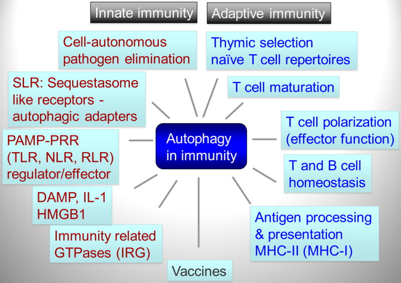
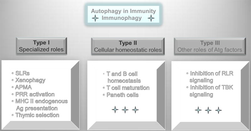
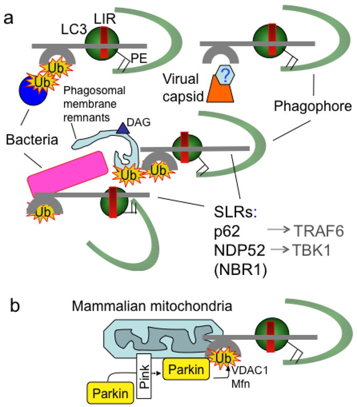
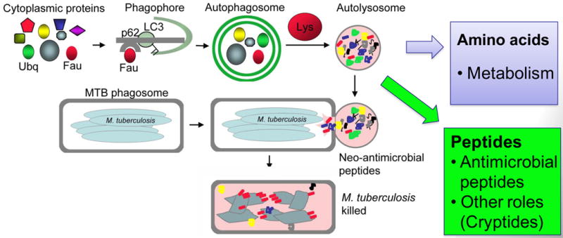
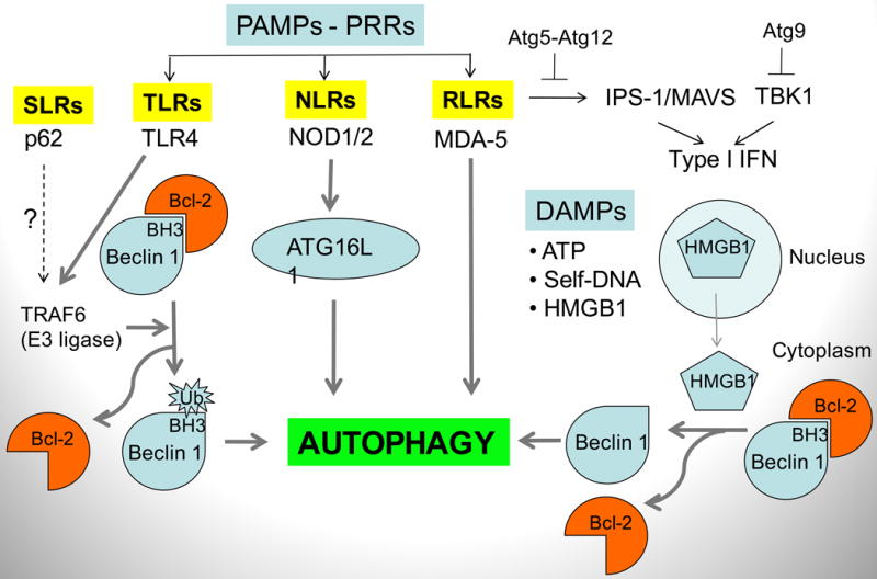
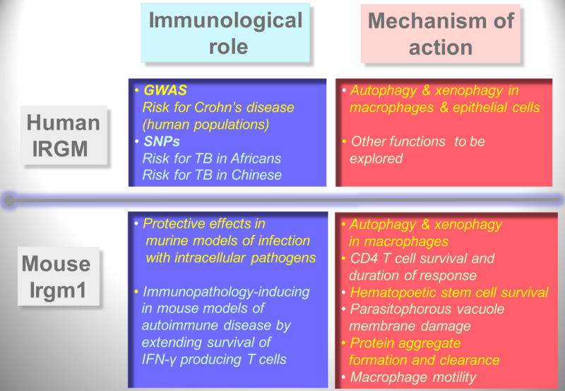
Autophagy was viewed until very recently primarily as a metabolic and intracellular biomass and organelle quality and quantity control pathway. It has now been recognized that autophagy represents a bona fide immunologic process with a wide array of roles in immunity. The immunologic functions of autophagy, as we understand them now, span both innate and adaptive immunity. They range from unique and sometimes highly specialized immunologic effectors and regulatory functions (referred to here as type I immunophagy) to generic homeostatic influence on immune cells (type II immunophagy), akin to the effects on survival and homeostasis of other cell types in the body. As a concept-building tool for understanding why and how autophagy is intertwined with immunity, it is useful to consider that the presently complex picture has emerged in increments, starting in part from the realization that autophagy acts as an evolutionarily ancient microbial clearance mechanism defending eukaryotic cells against intracellular pathogens. In this review, we build a stepwise model of how the core axis of autophagy as a cell-autonomous immune defense against microbes evolved into a complex but orderly web of intersections with innate and adaptive immunity processes. The connections between autophagy and conventional immunity systems include Toll-like receptors, Nod-like receptors, RIG-I-like receptors, damage-associated molecular patterns such as HMGB1, other known innate and adaptive immunity receptors and cytokines, sequestasome (p62)-like receptors that act as autophagy adapters, immunity-related GTPase IRGM, innate and adaptive functions of macrophages and dendritic cells, and differential effects on development and homeostasis of T- and B-lymphocyte subsets. The disease contexts covered here include tuberculosis, infections with human immunodeficiency virus and other viruses, Salmonella, Listeria, Shigella, Toxoplasma, and inflammatory disorders such as Crohn's disease and multiple sclerosis.


Figures

Fig. 1
Fig. 1. Autophagy plays multiple roles in innate and adaptive immunity
Autophagy functions within the innate immunity (red letters), adaptive immunity (blue letters), and their intersection (black font; e.g. vaccines). SLR, p62/Sequestasome like receptors that also function as autophagy adapters. PAMP, pathogen-associated molecular patterns. PRR, pattern recognition receptors. TLR, Toll-like receptors. NLR, Nod-like receptors. RLR, RIG-I-like receptors. DAMP, danger-associated molecular patterns. IRG, immunity-related GTPases.
Fig. 2
Fig. 2. Autophagy pathway in mammalian cells
I. Phase I (initiation): mTOR (mammalian target of rapamycin)-dependent and mTOR-independent pathways can induce autophagy. mTOR works via Ulk1 and 2 (mammalian Atg1 orthologs) that in turn directs (most likely on the ER) appropriate protein and lipid transactions to allow formation of a nascent pre-phagophore (1) possibly corresponding to the well defined structure in yeast termed pre-autophagosomal structure (or phagophore assembly site; PAS). The pre-phagophore in mammalian cells may emerge from an ER structure termed omegasome (Ω). Contributions of the secretory pathway [ER-Golgi-trans Golgi network (ER-G-TGN)], plasma membrane (PM)-derived organelles and mitochondria (MT) have also been reported: PM, acts possibly as additional source of lipid bilayers or a source of regulatory effects within the endosomal system with which autophagosomes merge during maturation; MT may contribute as a major source of phosphatidylethanolamine (PE) where phosphatydilserine (PS) decarboxylase is located on the MT inner membrane to generate PE from PS for conjugation of LC3 (LC3-II) and as a source of regulatory reactive oxygen intermediates that may promote autophagosome formation; ER-G-TGN contributions are probably complex and affecting the process directly and indirectly at multiple stages. Another major system regulating autophagy is the phosphatidylinositol 3-kinase hVPS34, which generates phosphatidylinositol 3-phopshate (PI3P). DFCP is a protein with two FYVE (PI3P-binding) domains and serves as a marker for the merger of the hVPS34/PI3P systems and TOR/Ulk at the point of autophagy initiation at the ER. hVPS34 is in a complex (often referred to as complex I) with multiple proteins, with Beclin 1 being key for autophagy altogether, and mAtg14 being key specifically for autophagy initiation. WIPI1 and 2 (mammalian Atg18) bind to PI3P and likely facilitate anteretrograde (and retrograde for recycling) membrane and protein flow to the nascent phagosomes. PI3P phosphatases, such as Jumpy/MTMR14, modulate progression of autophagy by tempering PI3P-dependent processes. II. Phase II (elongation and closure): Autophagic membrane is elongated to from a structure known as sensu stricto phagophore (or isolation membrane) based on two conjugation systems: (a) Atg5-Atg12 in complex with Atg16L, which acts as an E3 enzyme (nomenclature borrowed form the ubiquitination system) topologically restricting and enzymatically facilitating (b) conversion of LC3 to LC3-II (lipidated at the C-terminus by PE). Atg16 is a Crohn's disease risk locus in human populations and has also been found in complexes with clathrin at the plasma membrane (not shown). Elongating phagophore, also known as isolation membrane (2) wraps around its targets and eventually closes to form a sealed double membrane organelle known as sensu stricto autophagosome (3). III. Phase III (maturation). Autophagosomes fuse with late endosomal and lysosomal organelles or intermediates in a process dependent on hVPS34 (in complex with Beclin 1 and UVRAG; referred to as complex II). This process is also subject to inhibition/modulation by PI3P phosphatases such as Jumpy/MTMR14. The fusion with lysosomal organelles heralds the dissolution of the inner membrane and formation of autolysosomes, where the degradation of the captured material occurs.
Fig. 3
Fig. 3. Three types of autophagy intersections with immune processes
Type I immunophagy, encompasses specialized roles of autophagy in capturing, processing, or delivering microbes or microbial or endogenous immunologically active molecules. SLRs (p62/sequestasome-like receptors) that serve as bridges between microbial targets and autophagosomes. Xenophagy, process of direct capture and destruction of microbes in autolysosomes. APMA, autophagic activation of macrophages (a term describing the cumulative state of immunological activities and processes in macrophages induced for autophagy). PRR, pattern recognition receptors. Type II immunophagy refers to the role of autophagy in controlling cellular viability and general functionality of immune cells in ways that are not different than effects in all other cell types (e.g. neurons). Type III processes are those that are affected by isolated Atg factors but are not dependent on the execution of the entire autophagic pathway, as described in Fig. 2. For example, Atg5-Atg12 has been implicated in inhibition of RIG-I-like receptor (RLR) signaling, whereas Atg9 has been implicated in negatively regulating TBK1 as it contributes to type I interferon secretion.
Fig. 4
Fig. 4. SLRs (p62/sequestasome-like receptors) capture microbes or associated host structures for delivery to nascent autophagic organelles
(A). Capture of microbes (bacteria, viral components/capsid) or parasitophorous vacuole membrane remnants by SLRs for autophagic degradation. (B). Capture of terminally depolarized/stressed/damaged mitochondria by SLRs for autophagic removal (“mitophagy”). SLRs include p62 sequestasome, NDP52, NBR1. All of these molecules serve as autophagic adapters by the virtue of interacting with LC3 (one of mammalian Atg8s) or other mammalian Atg8 paralogs, often through a motif termed LIR (LC3 interaction region, with a variation of the ‘WXXL’ motif: D/EW/FE/DXLI/V). SLRs also interact with proinflammatory signaling factors such as TRAF6 and TBK1. Ub, ubiquitin tags recognized by ubiquitin binding regions (e.g. UBA in p62 and NBR1 or zinc finger in NDP52). DAG (triangle), diacylglicerol, a lipid tag found on microbe-harboring phagosomal membranes as a signal for autophagic degradation that may act independently of ubiquitin tags (Ub). Hexagon labeled ‘?’, tags associated with viral capsids have not been identified. Green semicircle, phagophore. PE, phosphatidylethanolamine. Parking, ubiquitin E3 ligase recruited to stressed mitochondria by Pink, with ubiquitination of VDAC1 (a component of mitochondrial permeability transition pore and the most abundant mitochondrial outer membrane protein) and Mfn (mitofusin, a GTPase that controls mitochondrial dynamics and fusion). Note the striking similarity between removal of intracellular microbes (a) and mitochondria (b) by SLRs, possibly reflecting ancient evolutionary relationships since mitochondria evolved from α-proteobacterial (Rickettsia-like microorganisms) symbionts.
Fig. 5
Fig. 5. Autophagy generates neoantimicrobial peptides
Autophagy has access to a variety of cytoplasmic proteins, such as FAU (a precursor for a ribosomal protein rpS30) and ubiquitin. Once sequestered into autophagosomes by an SLR, these proteins are subjected to proteolysis in autolysosomes. It is usually considered that proteins are degraded to amino acids and that amino acids are exported to the cytosol for nutritional purposes. However, autophagic organelles contain many proteolytic peptide intermediates, which when delivered by fusion with organelles (e.g. phagosomes) containing microbes (e.g. M. tuberculosis) act as antibacterial peptides. These have been termed neoantimicrobial peptides. However, the peptide mixture generated in autophagic organelles may harbor additional biological and signaling functions associated with potentially released or excreted peptides (termed cryptides).
Fig. 6
Fig. 6. Autophagy effectors and regulatory functions in the context of pattern recognition receptors (PRRs), pathogen-associated molecular patterns (PAMPs) and danger-associated molecular patterns (DAMPs)
Autophagy intersects with innate immunity at the levels of PAMPs and PRRs in all innate immunity receptor categories: TLRs (Toll-like receptors), NLRs (Nod-like receptors), RLRs (RIG-I-like receptors). It also interacts with molecules signaling tissue damage (DAMP), such as ATP, HMGB1, chromatin/DNA complexes, released from damaged cells. Mechanisms: TLR4 recruits TRAF6 that acts as an ubiquitin E3 ligase, ubiquitinating a Lys residue within the BH3 domain of Beclin 1, thus dissociating Bcl-2, an inhibitor of Beclin 1 function in promoting autophagy. Nod2 is in complexes with Atg16L1 and affects autophagy as well as Atg16L1 recruitment to the point of microbial entry into the cell (not shown). MDA-5 induces autophagy, shown in the context of cell death in malignancies. HMGB1 (high-mobility group protein B1; a non-histone component of eukaryotic chromatin) is an archetypical DAMP/alarmin released form the nucleus into the cytosol and in dying cells (mostly during necrosis) is released extracellularly. In the extracellular milieu, it signals through different receptors in tissue repair (when not complexed with microbial products such as lipopolysaccharide (LPS) or alarmin cytokines such as IL-1β) or in anti-microbial inflammation (when complexed with LPS or IL-1β). In the cytosol, HMGB1activates autophagy by binding to Beclin 1 and dissociating its inhibitory partner Bcl-2. Atg gene products (Atg5, Atg9, and Atg12) have been implicated in negative regulation of proinflammatory responses leading to type I interferon (IFN) production. It may be a reflection of type III non-autophagic functions of Atg genes, but it can be also viewed as a negative feedback mechanism to inhibit pro-inflammatory reactions induced in part through help of autophagy and Atg factors.
Fig. 7
Fig. 7. Summary of the known roles of human immunity related GTPase (IRG) IRGM and its murine ortholog Irgm1
Roles, left boxes; mechanisms, right boxes. Humans have one sensu stricto IRG gene, IRGM. Mouse has 22 sensu stricto IRG genes, of which Irgm1 is believed to be orthologous to human IRGM, via the IRGM9 evolutionary intermediate gene in pre-simians. During primate evolution, IRGM has been pseudogenized (by Alu repeat insertion and termination codon mutations) but then reconstituted in anthropoids (humans, chimpanzees, and gorillas) under the expression control by an endogenous retroviral element ERV9. Murine Irgm1 is under control by IFN-γ, whereas the human gene IRGM is not controlled by IFN-γ but is needed for IFN-γ-induced autophagy. GWAS, Genome-wide association studies. SNPs, single nucleotide polymorphisms. TB, tuberculosis. Listed are findings from human population studies for IRGM as a risk locus for Crohn's disease (a high incidence form of inflammatory bowel disease) and a risk locus tuberculosis (two different SNPs) thus far identified in African and Chinese populations. Not shown is that GWAS studies have linked another autophagy gene, ATG16L1, with Crohn's disease predisposition. Note that many of the functions assigned to the murine Irgm1 fit directly or indirectly with autophagy or autophagy-associated processes.

References

    1. Deretic V. Autophagy in innate and adaptive immunity. Trends Immunol. 2005;26:523–528. - PubMed
    1. Munz C. Enhancing immunity through autophagy. Annu Rev Immunol. 2009;27:423–449. - PubMed
    1. Deretic V, Levine B. Autophagy, immunity, and microbial adaptations. Cell Host Microbe. 2009;5:527–549. - PMC - PubMed
    1. Levine B, Deretic V. Unveiling the roles of autophagy in innate and adaptive immunity. Nat Rev Immunol. 2007;7:767–777. - PMC - PubMed
    1. Gutierrez MG, Master SS, Singh SB, Taylor GA, Colombo MI, Deretic V. Autophagy is a defense mechanism inhibiting BCG and Mycobacterium tuberculosis survival in infected macrophages. Cell. 2004;119:753–766. - PubMed

Publication types