Review
. 2011 Mar;240(1):252-68.
doi: 10.1111/j.1600-065X.2010.00984.x.

Mycobacterium tuberculosis and the intimate discourse of a chronic infection

Affiliations
Review

Mycobacterium tuberculosis and the intimate discourse of a chronic infection

David G Russell. Immunol Rev. 2011 Mar.

Abstract

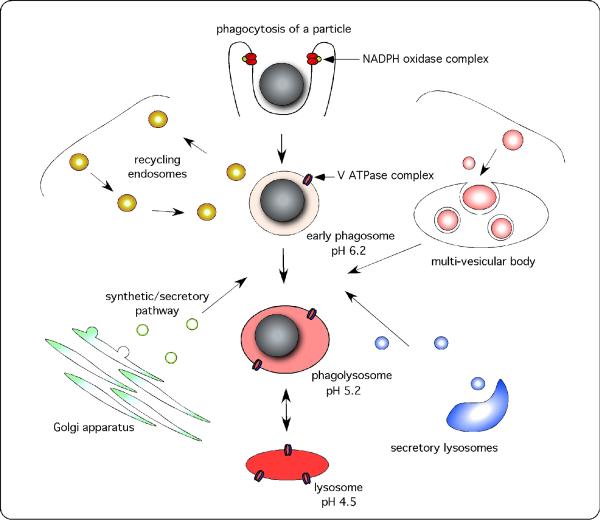
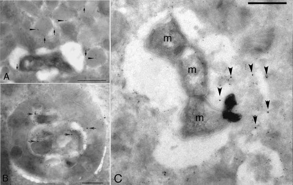
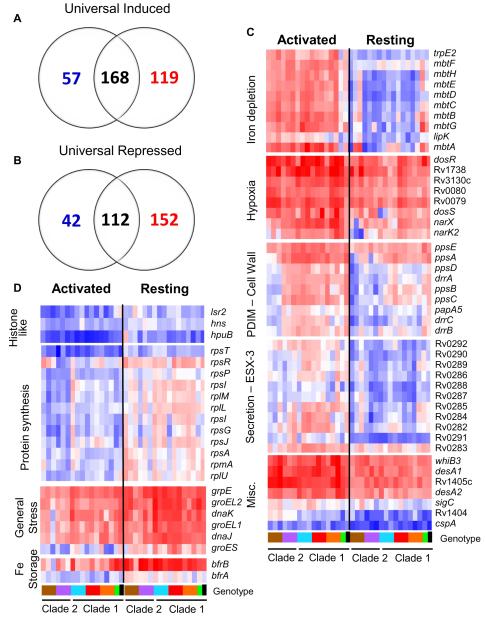
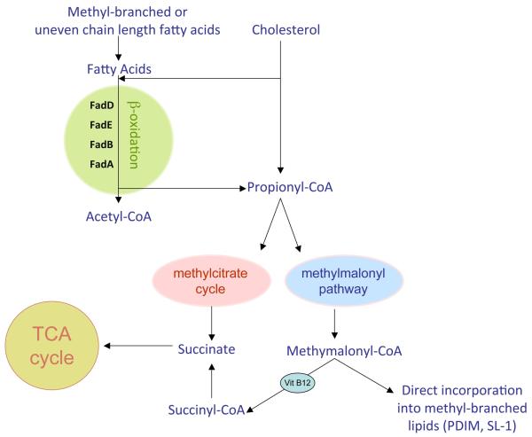
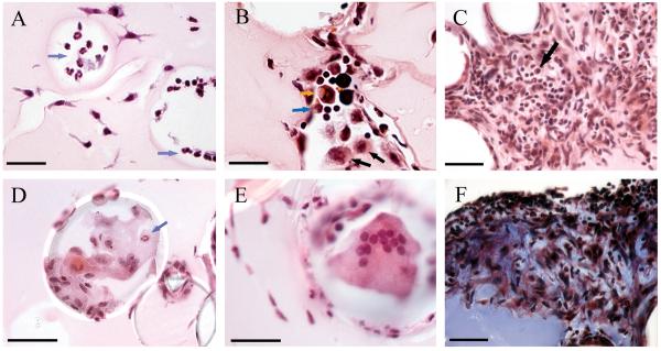
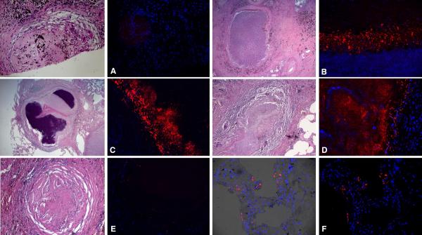
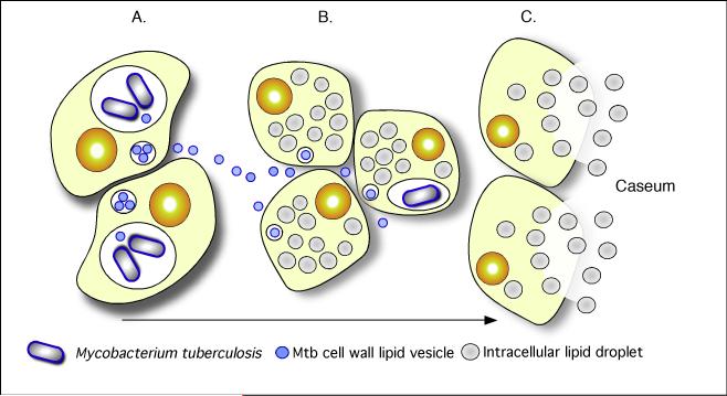
Mycobacterium tuberculosis is an extremely successful pathogen that demonstrates the capacity to modulate its host both at the cellular and tissue levels. At the cellular level, the bacterium enters its host macrophage and arrests phagosome maturation, thus avoiding many of the microbicidal responses associated with this phagocyte. Nonetheless, the intracellular environment places certain demands on the pathogen, which, in response, senses the environmental shifts and upregulates specific metabolic programs to allow access to nutrients, minimize the consequences of stress, and sustain infection. Despite its intracellular niche, Mycobacterium tuberculosis demonstrates a marked capacity to modulate the tissues surrounding infected cells through the release of potent, bioactive cell wall constituents. These cell wall lipids are released from the host cell by an exocytic process and induce physiological changes in neighboring phagocytes, which drives formation of a granuloma. This tissue response leads to the generation and accumulation of caseous debris and the progression of the human tuberculosis granuloma. Completion of the life cycle of tuberculosis requires damaging the host to release infectious bacteria into the airways to spread the infection. This damage reflects the pathogen's ability to subvert the host's innate and acquired immune responses to its own nefarious ends.


Figures

Fig. 1
Fig. 1. During maturation, the phagosome forms transient interactions with a wide range of intracellular organelles
Phagosome maturation refers to the process of phagosome remodeling through a series of independent events, following its formation at the surface of the phagocyte, and culminating in the complete fusion of the phagosome with the lysosome. Following engagement of phagocytic receptors, an area of the surface is remodeled around the particle, forming the phagocytic cup. Some protein complexes, such as the NADPH oxidase complex, can be recruited and activated prior to phagosomal closure, facilitating a rapid antimicrobial response at the cell surface. The phagosome, once closed, becomes increasingly more acidic, through the accumulation of V ATPases that pump protons into the compartment, and hydrolytically competent, through the acquisition of lysosomal enzymes. This process is marked by transient fusion events with multiple intracellular organelles including the recycling endosomal machinery, the synthetic–secretory apparatus including the endoplasmic reticulum, secretory lysosomes, and multi-vesicular bodies, which may include the MIIC compartment and/or the autophagosome. Finally, the phagosome fuses with pre-existing, dense lysosomal bodies and equilibrates to a pH of 4.5-5.0.
Fig. 2
Fig. 2. Spectrofluorometric measurement of the superoxide burst generated by bone marrow-derived murine macrophages following internalization of IgG-coated beads
An illustration of the assay that we have developed for measuring the superoxide burst within the phagosome. Murine macrophages were fed IgG-coated particles labeled with the oxidation sensitive fluorochrome dihydrohexafluorofluorescein, and the calibration dye Alexa-594. Both wildtype and macrophages deficient in the p91 subunit of the NADPH oxidase were examined in their resting and LPS-activated states, and the increase in fluorescence measured by spectrofluorometer. Activation of the macrophage with LPS enhanced the intensity but not the duration of the burst. Reproduced from (13).
Fig. 3
Fig. 3. Immunoelectronmicrograph from a human alveolar macrophage isolated from a TB patient in Malawi
Following isolation the cells were incubated with the pH-indicator DAMP, which accumulates in acidic compartments, for 60 minutes prior to fixation for electron microscopy. The sample was sectioned and probed with anti-DNP (12 nm gold particles) and anti-LAMP 1 (6 nm gold particles). The acidified lysosomes label strongly (arrowhead), while the Mtb-containing vacuoles show minimal to no labeling indicating that their pH is considerably higher than that of the neighboring lysosomes. Reproduced from (38).
Fig. 4
Fig. 4. Measurement of hydrolytic activity in phagosomes using fluorogenic substrates
(A). Cysteine proteinase substrate hydrolysis profiles for IgG-coated bead containing phagosomes. The beads were carrying the cysteine proteinase substrate Biotin-LC-Phe-Arg)2-Rhodamine 110, which increased fluorescence following the removal of the Phe-Arg quenching peptides. Manipulation of hydrolytic rates was achieved with inhibitors Concanamycin A (100 nM), W7 (15 μM) and Leupeptin (100 μg/ml), both of which reduced the rate of hydrolysis within the phagosome. (B). Triglyceride substrate hydrolysis profiles for IgG-coated bead containing phagosomes. These beads were carrying the lipase substrate 1-trinitrophenyl-amino-dodecanoyl-2-pyrenedecanoyl-3-O-hexadecyl-sn-glycerol. In contrast to proteolysis, blocking acidification of the phagosome increased its lypolytic activity. Traces were generated using the equation FU=R110/AF594 (where FU=Arbitrary Fluorescent Units, R110=Real time Rhodamine 110 fluorescence, AF594=Starting Alexa Fluor 594 fluorescence) and averaged over two experiments. Measurements were taken every second for 30 minutes. Reproduced from (14)
Fig. 5
Fig. 5. Immunoelectron microscopy of Mtb-infected macrophages probed with antibodies against ubiquitinated proteins
(A). Resting, Mtb-infected macrophages show ubiquitin label (arrowheads) in dense, LAMP1-positive (arrows) lysosomes. (B). In cells in which autophagy has been induced, the ubiquitin label can be seen in multivesicular structures that are reminiscent of autophagosomes. (C). In Mtb-infected cells in which autophagy has been induced the ubiquitinated material (arrows) can be seen in the Mtb-containing phagosomes. Reproduced from (70).
Fig. 6
Fig. 6. The ‘universal’ intracellular transcriptome of Mtb
The MTC universal intracellular transcriptome: identification of genes with conserved expression and regulation inside macrophage phagosome across global panel of MTC clinical isolates. Global gene expression profiles of 17 MTC strains 24h post-infection of resting and activated macrophages were determined by microarray. Normalized expression ratios for each strain were determined by comparison of RNA from intracellular bacteria versus control bacteria of the same strain treated identically except for phagocytosis by macrophages. This serves to identify relative responses to phagosomal cues rather than inherent strain-dependent differences in gene expression. (A-B) Venn diagrams showing activation-dependent (red=activated, blue=resting) and independent (black) genes with conserved expression patterns across clinical isolates. ‘Universal genes’ were selected based on trending up or down in all strains (up or down >1.2-fold in 15 of 17 strains) and significant induction or repression in >50% of strains (up or down >1.5X in 8 of 17 strains) in each macrophage type. The starting gene list for this analysis included only genes flagged as present in the majority of samples from both macrophage types. (C) Select genes with higher expression levels in activated versus resting macrophages conserved across all or most clinical isolates. Represents subset of activation-dependent genes identified by one-way ANOVA analysis of all intracellular transcription profiles (Benjamini and Hochberg False Discovery Rate p<0.01). (D) Select genes with higher expression in resting versus activated macrophages. See (C) above for analysis description. Refer to Fig. 1B for genotype color bar definition (black box denotes reference strain CDC1551). Reproduced from (84).
Fig. 7
Fig. 7. Outline of the pathways relevant to C3 metabolism
The metabolism of cholesterol, methyl-branched fatty acids and odd-chain length lipids will raise the intracellular levels of the C3 compounds propionate or propionyl-CoA, which Mtb finds highly toxic. The bacterium has developed three different strategies to detoxify propionyl-CoA. Isocitrate lyase activity has been suborned to fulfill the function of methylcitrate lyase in the last step of the methyl citrate cycle to generate the TCA cycle intermediate succinate. The methylmalonyl pathway has also been mobilized to metabolize propionyl-CoA to produce succinyl-CoA via the VitB12-dependent activity of methylmalonyl-CoA mutase. Finally, intermediates from the methylmalonyl pathway can be incorporated directly into the abundant, methyl-branched lipids of the bacterial cell wall, such as PDIM and SL-1. Reproduced from (129).
Fig. 8
Fig. 8. Mtb-derived cell wall lipids transfer from infected to uninfected macrophages
Transfer of mycobacterial material from BCG-infected macrophages to uninfected bystanders following overnight incubation. BMMØ infected with Texas Red hydrazide-labeled BCG were incubated with an equal number of uninfected macrophages labeled with chloromethylfluorescein diacetate. Uninfected bystander cells (green fluorescence) acquired Texas Red-labeled mycobacterial material from BCG-infected macrophages. Reproduced from (76).
Fig. 9
Fig. 9. Cells associated with BCG lipid-coated beads
Matrices containing BMMØ and BCG lipid-coated beads were injected i.p. and recovered at 14 h (A), 4 days (B) and 12 days (C - F). (A): Neutrophils (blue arrows) were present in spaces that had been occupied by beads after 14 hr. Scale bar, 35 mm. (B): A diverse infiltrate of macrophages (black arrows), neutrophils (blue arrow), eosinophils (orange arrow) and lymphocytes (small cells with little cytoplasm) accumulated at beads at 4 days. Scale bar, 25 mm. (C): A dense cellular infiltrate composed of mononuclear and polymorphonuclear leukocytes and lymphocytes, including plasma cells (black arrow) formed at 12 days. Infiltrates were especially florid in spaces between lipid-coated beads. Scale bar, 35 mm. (D): Epithelioid macrophages and occasional neutrophils (blue arrow) typically adhered to beads. Scale bar represents 50 mm. (E): Multi-nucleated giant cell in association with a bead at 12 days. Scale bar, 35 mm. (F): Fibrotic material (dark blue-staining material) was deposited in the dense aggregates of leukocytes at 12 days. The uniformly staining blue material in the lower left corner of the panel is the collagen in the Matrigel. Scale bar, 35 mm. Representative H & E-stained sections (A – E) and a trichrome-stained section (F) are shown. Reproduced from (97).
Fig. 10
Fig. 10. ADFP expression in human TB granulomas
Immunofluorescence signals were obtained for each granuloma, and a representative image (right) and the corresponding region from an H&E stained slide (left, boxed) are shown. Nuclei are seen in blue and antigens in red. (A). Nascent granulomas exhibit weak ADFP expression. (B). Caseous granulomas stain strongly for ADFP. (C). Fibrocaseous granulomas also label strongly for ADFP expression. (D). The caseous center also has intense ADFP expression, together with nuclear debris. E. Resolved granulomas exhibit low levels of ADFP labeling. F. Control normal lung parenchyma shows ADFP expression in pneumocytes and alveolar macrophages (the left image is a merged image with bright field). Scale bar is 50 mm. Reproduced from (102).
Fig. 11
Fig. 11. A model illustrating the linkages between Mtb-infection, foam cell formation and accumulation of caseum in the human TB granuloma
(A). Intracellular Mtb bacilli synthesize and release cell wall components inside their host cells. We have demonstrated previously that these lipids accumulate in the internal vesicles in multi-vesicular bodies, which are exocytosed from the cell in vesicular form (B). Because of the release of these vesicles, both infected and uninfected macrophages are exposed to cell wall mycolates and induced to form foamy macrophages, as illustrated in Fig. 7. The foamy macrophages have been shown to support the maintenance and growth of persistent bacteria (C). We now propose that these cells die via an inflammatory, necrotic process and release their lipid droplets into the extracellular milieu within the granuloma. As a result of the fibrotic capsule, the human granuloma is an enclosed, isolated structure with minimal vasculature. The enclosed nature of the human granuloma leads to accumulation of necrotic debris as caseum. In this model, this process is an integral part of the pathology that leads to active disease and transmission. Reproduced from (102).

Similar articles

Cited by

References

    1. Russell DG, Barry CE, 3rd, Flynn JL. Tuberculosis: what we don’t know can, and does, hurt us. Science. 2010;328:852–856. - PMC - PubMed
    1. Cardoso CM, Jordao L, Vieira OV. Rab10 regulates phagosome maturation and its overexpression rescues Mycobacterium-containing phagosomes maturation. Traffic. 2010;11:221–235. - PubMed
    1. Philips JA, Porto MC, Wang H, Rubin EJ, Perrimon N. ESCRT factors restrict mycobacterial growth. Proc Natl Acad Sci USA. 2008;105:3070–3075. - PMC - PubMed
    1. Roberts EA, Chua J, Kyei GB, Deretic V. Higher order Rab programming in phagolysosome biogenesis. J Cell Biol. 2006;174:923–929. - PMC - PubMed
    1. Roberts EA, Deretic V. The Mycobacterium tuberculosis phagosome. Methods Mol Biol. 2008;445:439–449. - PubMed

Publication types