. 2011 Jan 18;8(1):2.
doi: 10.1186/2045-8118-8-2.

Fluids and barriers of the CNS: a historical viewpoint

Affiliations

Fluids and barriers of the CNS: a historical viewpoint

Shane A Liddelow. Fluids Barriers CNS. .

Abstract

Tracing the exact origins of modern science can be a difficult but rewarding pursuit. It is possible for the astute reader to follow the background of any subject through the many important surviving texts from the classical and ancient world. While empirical investigations have been described by many since the time of Aristotle and scientific methods have been employed since the Middle Ages, the beginnings of modern science are generally accepted to have originated during the 'scientific revolution' of the 16th and 17th centuries in Europe. The scientific method is so fundamental to modern science that some philosophers consider earlier investigations as 'pre-science'. Notwithstanding this, the insight that can be gained from the study of the beginnings of a subject can prove important in the understanding of work more recently completed. As this journal undergoes an expansion in focus and nomenclature from cerebrospinal fluid (CSF) into all barriers of the central nervous system (CNS), this review traces the history of both the blood-CSF and blood-brain barriers from as early as it was possible to find references, to the time when modern concepts were established at the beginning of the 20th century.


Figures
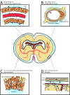
Figure 1
Figure 1
Illustration of the sites of brain barriers in the developing and adult brain. A. The blood-CSF barrier. A barrier between choroid plexus blood vessels and the CSF. The blood vessels in the choroid plexus are fenestrated and form a non-restrictive barrier (dotted arrows). The choroid plexus epithelial cells (cpecs) are joined by functional tight junctions towards their apical surface that stop the movement of molecules (arrows). B. The blood-brain barrier. A barrier between the lumen of cerebral blood vessels and the brain parenchyma. The endothelial cells have luminal tight junctions (arrow) that form the physical barrier stopping the movement of molecules out of the vasculature. Astrocytic endfeet are in close association of the cerebral blood vessels and form what is known as the 'neurovascular unit'. The endfeet are not necessary for blood-brain barrier integrity. C. The inner CSF-brain barrier, present only during early development. A barrier between the CSF and the brain parenchyma. The neuroependymal cells lining the ventricular wall (orange) are connected by 'strap junctions' [2], halting the exchange of large molecules such as proteins between the CSF and brain (arrows), but not of smaller molecules like sucrose. This barrier is not present in the adult brain due to a loss of strap junctions. There is no restriction of movement at this time. D. The outer CSF-brain barrier. A barrier between the CSF-filled subarachnoid space (sas) and overlying structures. The blood vessels in this area are fenestrated and provide little by way of a barrier, but the outer cells of the arachnoid membrane (arach) are connected by tight junctions. Abbreviations: arach, arachnoid membrane; cpec, choroid plexus epithelial cells; dura, dura mater; nu., nucleus; pia, pia mater; sas, subarachnoid space. Adapted with permission from [4].
Figure 2
Figure 2
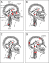
The evolution of our understanding of CSF production and pathway in relation to brain spaces. A. Cladius Galenus' (Galen, 129 - 219AD) concept of the CSF pathway. In his writings Galen describes a lateral ventricular choroidal origin (1) and exit through the fourth ventricle to the spinal canal (2). He also erroneously describes movement of the fluid across the cribriform plate into the nasal cavity (3) and across the infundibulum to the palate (4). B. Albrecht von Haller's (1708 - 1777AD) CSF pathway correctly stated the origin of CSF from the ventricles (1), with exit from the fourth ventricle (2) and down the spinal canal for venous absorption. This early description was essentially correct. C. François Magendie's (1783 - 1855AD) concept of the CSF pathway was exactly opposite to the system of both von Haller and Galen. D. The modern description of the CSF pathway. CSF is produced by the choroid plexuses (1) from where it moves from the lateral ventricles into the third and fourth ventricles (2). It then flows across the surface of the brain (3) and down the spinal canal (moving from the back to front (3) of the canal). CSF is then reabsorbed by the arachnoid granulations (4) back into the blood stream. The arachnoid villi are projections from the arachnoid layer of the meninges that connect with veins via the venous sinus. Absorption into lymphatics also occurs (not shown). Abbreviations: 3V, third ventricle; 4V, fourth ventricle; LV, lateral ventricle.
Figure 3
Figure 3
Decorated letters from Vesalius' De Humani Coporis Fabrica (1543). A. The act of removing the head of cadavers prior to dissection can be seen, as cherubs set to the head of a man with a bone saw. B. Once decapitated, the subjects (human or beast) were hung to drain the blood from the body. This enabled a much 'cleaner' visualisation of the internal organs. C/D. At the same time as public dissections of human subjects were occurring, the dissections of beasts was also conducted. This increased the availability of subjects, but also allowed a better comparison to the works of the ancients who worked mostly on animals. In C, the cherubs can be seen dissecting a boar, while another looks on, reading from a book (possibly Galen's). E/F. The difficulty in obtaining human subjects was such that at the time of Vesalius' bodies were often obtained via grave robbing (E) or by taking the bodies of executed criminals (F). All images are reproduced by gracious permission of Her Majesty The Queen, from the Royal Collection © 2010, Her Majesty Queen Elizabeth II.
Figure 4
Figure 4
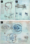
Sketches by Leonardo da Vinci on the anatomy of the brain. A. The layers of the scalp compared to an onion (1489). The earliest drawings by da Vinci on the ventricles of the brain show them to be connected to the eye and moving backwards, into the brain. In this drawing he also likens the meninges of the brain to the layers of an onion (left hand side of image). B. Studies of the eyes and brain (1508). Later studies by da Vinci on the neuroanatomy of man display a better understanding of the ventricles of the brain and of nerves permeating to peripheral areas. This increase in understanding is likely due to the use of wax casts made of the ventricular system of other 'lower' animals, such as the ox (see C). C. The cerebral ventricles of the brain of an ox (unknown date: 1508 - 1510). Here da Vinci describes the methodology of injecting warmed wax into the ventricular system of the ox, allowing it to cool, then visualising and sketching the mould that is made. Though not describing explicitly the use of bovine species, we can assume that da Vinci has by the presence of the bovine equivalent of the circle of Willis, the rete mirabilis, in the lower image. All images are reproduced by gracious permission of Her Majesty The Queen, from the Royal Collection © 2010, Her Majesty Queen Elizabeth II.
Figure 5
Figure 5
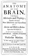
Recto Title Page of Humphrey Ridley's 1695 book 'The Anatomy of the Brain'. This test contains a comment on the 'tightness' of cerebral blood vessels, approximately 150 years before the experiments of the German scientists Ehrlich, Goldmann and Lewandowsky.
Figure 6
Figure 6
Illustration of early brain barrier experiments by Ehrlich and Goldmann. These early experiments elegantly demonstrated the compartmentalisation between the central nervous system (brain and spinal cord) and the peripheral organs. A. Trypan blue is delivered peripherally [86,88]. The dye does not penetrate any organs of the central nervous system, which both researchers suggested was due to the central nervous system having a lower affinity than other tissues. B. Trypan blue is injected into the brain [12]. The brain and spinal cord were stained, while the peripheral organs were not.

References

    1. Barcroft J, Barron D. Factors influencing the oxygen supply of the brain at birth. J Physiol USSR. 1938;24:43–55.
    1. Møllgård K, Balslev Y, Lauritzen B, Saunders NR. Cell junctions and membrane specializations in the ventricular zone (germinal matrix) of the developing sheep brain: a CSF-brain barrier. J Neurocytol. 1987;16:433–444. - PubMed
    1. Janzer RC, Raff MC. Astrocytes induce blood-brain barrier properties in endothelial cells. Nature. 1987;325:253–257. doi: 10.1038/325253a0. - DOI - PubMed
    1. Saunders NR, Knott GW, Dziegielewska KM. Barriers in the immature brain. Cell Mol Neurobiol. 2000;20:29–40. doi: 10.1023/A:1006991809927. - DOI - PMC - PubMed
    1. Daneman R, Zhou L, Kebede AA, Barres BA. Pericytes are required for blood-brain barrier integrity during embryogenesis. Nature. 2010;468:562–566. doi: 10.1038/nature09513. - DOI - PMC - PubMed