Comparative Study
. 2011 Mar 1;57(9):1081-92.
doi: 10.1016/j.jacc.2010.09.066.

Mechanisms of fractionated electrograms formation in the posterior left atrium during paroxysmal atrial fibrillation in humans

Affiliations
Comparative Study

Mechanisms of fractionated electrograms formation in the posterior left atrium during paroxysmal atrial fibrillation in humans

Felipe Atienza et al. J Am Coll Cardiol. .

Abstract

Objectives: The aim of this paper was to study mechanisms of formation of fractionated electrograms on the posterior left atrial wall (PLAW) in human paroxysmal atrial fibrillation (AF).

Background: The mechanisms responsible for complex fractionated atrial electrogram formation during AF are poorly understood.

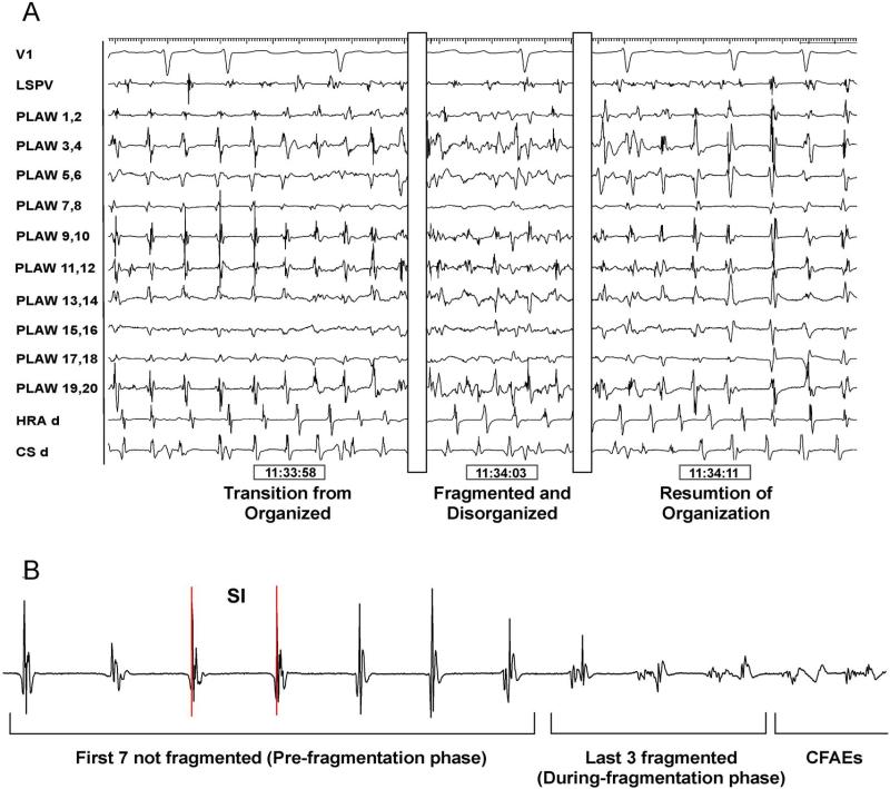
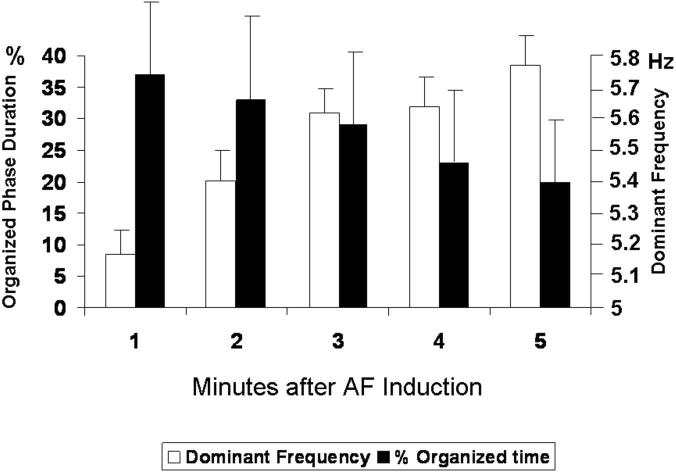
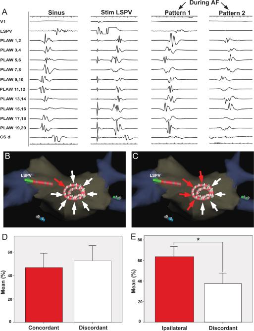
Methods: In 24 patients, we induced sustained AF by pacing from a pulmonary vein. We analyzed transitions between organized patterns and changes in electrogram morphology leading to fractionation in relation to interbeat interval duration (systolic interval [SI]) and dominant frequency. Computer simulations of rotors helped in the interpretation of the results.

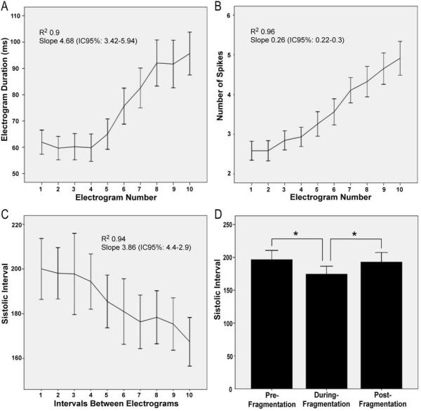
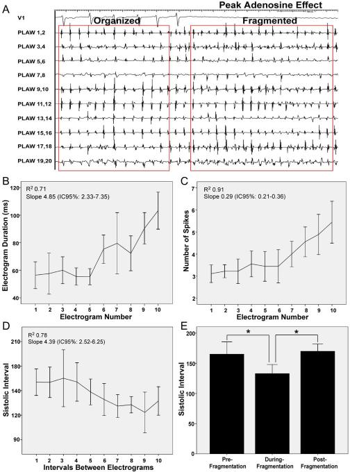
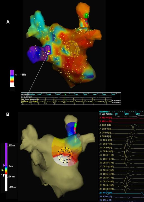
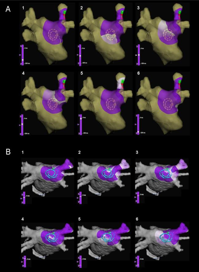
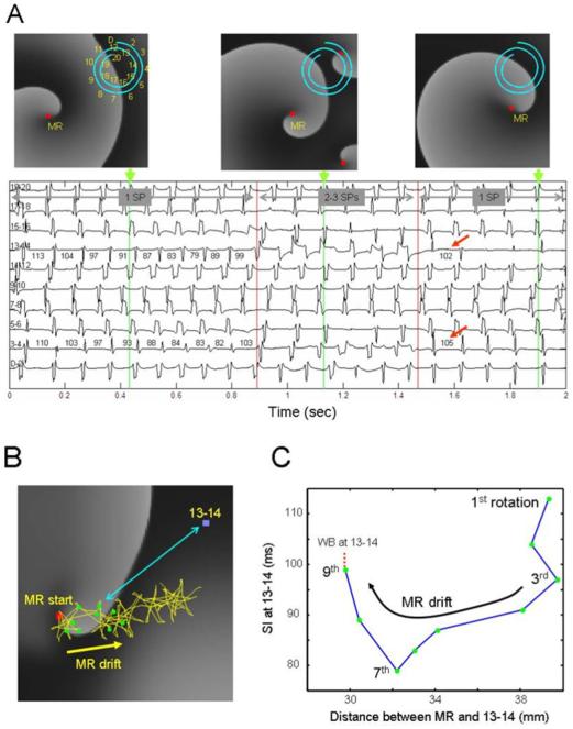
Results: Organized patterns were recorded 31 ± 18% of the time. In 47% of organized patterns, the electrograms and PLAW activation sequence were similar to those of incoming waves during pulmonary vein stimulation that induced AF. Transitions to fractionation were preceded by significant increases in electrogram duration, spike number, and SI shortening (R(2) = 0.94). Similarly, adenosine infusion during organized patterns caused significant SI shortening leading to fractionated electrograms formation. Activation maps during organization showed incoming wave patterns, with earliest activation located closest to the highest dominant frequency site. Activation maps during transitions to fragmentation showed areas of slowed conduction and unidirectional block. Simulations predicted that SI abbreviation that heralds fractionated electrograms formation might result from a Doppler effect on wave fronts preceding an approaching rotor or by acceleration of a stationary or meandering, remotely located source.

Conclusions: During induced AF, SI shortening after either drift or acceleration of a source results in intermittent fibrillatory conduction and formation of fractionated electrograms at the PLAW.


Figures

Figure 1
Figure 1. Transitions between organized and fragmented phases
A. Posterior left atrial wall (PLAW) intracardiac recordings using a 20 electrodes spiral catheter. From left o right, transitions from organized to fragmented and back to organized electrograms. B. Ten consecutive electrograms were analyzed prior to fragmentation: last 7 organized electrograms (pre-fragmentation phase) and first 3 electrograms showing fragmentation (during-fragmentation phase). CFAE, complex fractionated atrial electrograms; CS, coronary sinus; HRA, high right atrium; SI, systolic interval.
Figure 2
Figure 2. Time courses of organization and dominant frequency
Posterior left atrial wall temporal changes in the percent of organized phases duration (black bars) and dominant frequency (white) after induction.
Figure 3
Figure 3. Electrogram characteristics and systolic interval (SI) during transitions from organized to fragmentation (first 10 complexes)
A. Electrogram duration; B. Number of spikes. C. SI. D. Mean values of SI before and during fragmentation and after resumption of organization.
Figure 4
Figure 4. Adenosine infusion
A. Tracings during peak adenosine effect. Lead V1 and intracardiac electrograms recorded from spiral catheter at the posterior left atrial wall (PLAW). At peak adenosine effect (complete AV block) transition from organized to fragmented electrograms is observed, with simultaneous cycle length shortening. B, C and D. Electrogram duration, number of spikes and systolic interval (SI) during transitions from organized to fragmented electrograms (first 10 complexes). E. Mean values of SI before and during fragmentation, and after resumption of organization.
Figure 5
Figure 5. Organized patterns at the posterior left atrial wall
A. Left, activation patterns during sinus rhythm and during stimulation from the left superior pulmonary vein (LSPV). Right, two activation patterns were observed during atrial fibrillation (AF): pattern 1 resembled LSPV stimulation; pattern 2 was similar to sinus rhythm activation. B and D. During AF, concordant patterns of activation resembling LSPV stimulation from which AF was induced (red arrow originating at the pacing catheter) accounted for 46±27% of recording time when compared discordant patterns (white arrows). C and E. Organized patterns during AF resembling those obtained during pacing from any of the ipsilateral PVs (red arrows originating from the pacing PV side, superior and inferior) accounted for 63±24% of the organized time (p=0.025). CSd, distal coronary sinus.
Figure 6
Figure 6. Organized activation patterns in relation to dominant frequency sites location
A. Left atrial dominant frequency (DF) map (posterior view). White arrow points to highest DF site (10.8 Hz) at the left inferior pulmonary vein (LIPV) antrum. B. Posterior left atrial wall activation map during organized phase prior to fragmentation (right) shows an incoming wave pattern of activation progressing from closest to the highest DF site at the LIPV (left, white) to the right (purple-blue).
Figure 7
Figure 7. Snapshots of wave propagation at the posterior left atrial wall (PLAW) during transitions to fragmentation
(sequence 1–6): purple, unactivated regions; white, advancing activation. A. Reentrant circuit with a clockwise propagation around a pivoting point located to the right edge of the septo-pulmonary bundle. B. Activation breakthrough across a line of slow conduction, with activation of the PLAW in both superior and inferior directions, and final convergence on the anterior aspect of the right superior pulmonary vein antrum.
Figure 8
Figure 8. Simulation of a drifting rotor with peripheral wavebreaks (WB) in an atrial model
A. Snapshots of the model membrane voltage are at times indicated by the green markers. A 20 electrode catheter (D-20) shown on snapshots indicates locations of 10 pseudo-bipoles shown below maps. Red squares: Pivoting sites of rotors (i.e., singularity point; SP) Numbers on bipoles 3–4 and 13–14 indicate cycle length in ms. Horizontal gray arrows indicate episodes with a single mother rotor (MR, 1 SP), additional 2 short living rotors after ~0.9 sec (3 SPs) and their disappearance after ~1.5 sec (1 SP). Red arrows denote return of cycle length to pre-shortening phase. B. Trajectory of the drifting rotor's tip (yellow trace and arrow) superimposed on a snapshot of voltage at time=0. Red square, starting point of the drift; green dots, the location of the tip at the completion of each of 9 initial rotations; blue square, location of bipoles 13–14; double-headed blue arrow, distance between 13–14 bipoles and a sample position of the rotor's tip. C, Systolic Interval (SI) at bipoles 13–14, as a function of distance between the rotor's tip and the bipoles' location. As the drifting rotor gets closer to the bipoles, SI abbreviates due to Doppler shift, particularly after the 3rd rotation. After the 7th rotation, local conduction impairment at 13–14 increases SI with an eventual WB.

References

    1. Haïssaguerre M, Jaïs P, Shah DC, et al. Spontaneous initiation of atrial fibrillation by ectopic beats originating in the pulmonary veins. N Engl J Med. 1998;339:659–666. - PubMed
    1. Sanders P, Berenfeld O, Hocini M, et al. Spectral analysis identifies sites of high-frequency activity maintaining atrial fibrillation in humans. Circulation. 2005;112:789–797. - PubMed
    1. Oral H, Scharf C, Chugh A, et al. Catheter ablation for paroxysmal atrial fibrillation: segmental pulmonary vein ostial ablation versus left atrial ablation. Circulation. 2003;108:2355–2360. - PubMed
    1. Nademanee K, McKenzie J, Kosar E, et al. A new approach for catheter ablation of atrial fibrillation: mapping of the electrophysiologic substrate. J Am Coll Cardiol. 2004;43:2044–2053. - PubMed
    1. Di Biase L, Elayi CS, Fahmy TS, et al. Atrial Fibrillation Ablation Strategies for Paroxysmal Patients: Randomized Comparison Between Different Techniques. Circ Arrhythmia Electrophysiol. 2009;2:113–119. - PubMed

Publication types

MeSH terms