Review
. 2011 Apr;21(2):283-90.
doi: 10.1016/j.conb.2011.02.001. Epub 2011 Feb 23.

Structure and mechanism of glutamate receptor ion channel assembly, activation and modulation

Affiliations
Review

Structure and mechanism of glutamate receptor ion channel assembly, activation and modulation

Mark L Mayer. Curr Opin Neurobiol. 2011 Apr.

Abstract

Ionotropic glutamate receptors (iGluRs) are ligand gated ion channels that mediate excitatory synaptic transmission in the brain of vertebrates. A rapidly growing body of crystal structures for isolated iGluR extracellular domains, and more recently a full length AMPA receptor, combined with data from electrophysiological experiments and MD simulations, provides a framework that makes it possible to investigate the molecular basis for assembly, gating and modulation. These unprecedented advances in structural biology are constantly challenged by novel functional properties that emerge despite decades of functional analysis, and by a growing family of auxiliary proteins that modulate iGluR activity and assembly.


Figures

Figure 1
Figure 1
Domain organization in the GluA2 ion channel crystal structure (PDB 3KG23). (a) The four subunits in the tetramer assembly are colored blue, yellow, red and green; the molecular surface representation reveals key features of the structure, including its organization in layers, the cavity at the ATD and LBD interface, and the large size of the extracellular domains compared to the ion channel. (b) ATD dimer formed by chains A and B reveals a large separation of the LBD domains in this subunit pair; linkers which connect the layered domains are indicated by black arrows which draw attention to the different conformations of the LBD linkers in the A and B subunits. (c) LBD dimer formed by chains A and D reveals a large separation of the ATD domains in this subunit pair.
Figure 2
Figure 2
AMPA receptor assembly mechanisms revealed by synchronized protein expression and single particle EM analysis (from Ref. [•7]). (a) Schematic size exclusion chromatography profiles for elution of wild type GluA2 reveals a mixture of dimers and tetramers 20 hours after induction of expression, with an increase in tetramer and decrease in dimer populations 24 hours after induction; the lower panel shows chromatograms recorded at 24 hours for wild type GluA2 and the L483Y mutant which is trapped at the dimer stage of assembly. (b) Single particle EM analysis for wild type GluA2 dimers (top) with crystal structures for a GluA2 ATD dimer, and two GluA2 LBD monomers docked in the EM density map (bottom). (C) A similar analysis for the GluA2 L483Y dimer in which both the ATD and LBD layers contain dimer assemblies.
Figure 3
Figure 3
High resolution function and crystal structures for iGluR extracellular domains. (a) Tetramer assemblies of the kainate receptor GluK2 and GluK3 ATDs formed by a dimer of dimer assembly (from Ref. [25]). (b) iGluR loop 1 LBD structures superimposed using C coordinates for helix B revealing the elaborate structure in NMDA receptor subunits (from Ref. [25]). (c) Modulation of GluK1 outward current responses recorded at +60 mV by replacement of extracellular Na+ by Rb+ or Cs+. (d) The GluK2 M239D mutant retains activity in the presence of 100 μM Ca2+ and Mg2+ when Na is replaced by NMDG or Tris but is inhibited by 100 μM EDTA (from Ref. [48]). (e) Allosteric ion binding sites in the GluK1 LBD dimer assembly with alternate conformations for the Arg744 side chain and a 2:1 stoichiometry for Na+ and Cl (from Ref. [•46]). (f) MD simulation of the GluK2 ligand binding domain docked with glutamate; red spheres indicate the position of water molecules trapped in the ligand binding site in the crystal structure; the red mesh shows MD trajectories for individual water molecules; W1, W2, W2 and W4 freely exchange with other on a ns time scale (from Ref. [64]).

References

    1. Hollmann M. Structure of ionotropic glutamate receptors. In: Jonas P, Monyer H, editors. Ionotropic glutamate receptors in the CNS. Springer-Verlag; 1999. pp. 3–98.
    2. Handbook of experimental pharmacology. Vol. 141
    1. Traynelis SF, Wollmuth LP, McBain CJ, Menniti FS, Vance KM, Ogden KK, Hansen KB, Yuan H, Myers SJ, Dingledine R. Glutamate receptor ion channels: structure, regulation, and function. Pharmacol Rev. 2010;62:405–496. - PMC - PubMed
    1. Mayer ML. Glutamate receptors at atomic resolution. Nature. 2006;440:456–462. - PubMed
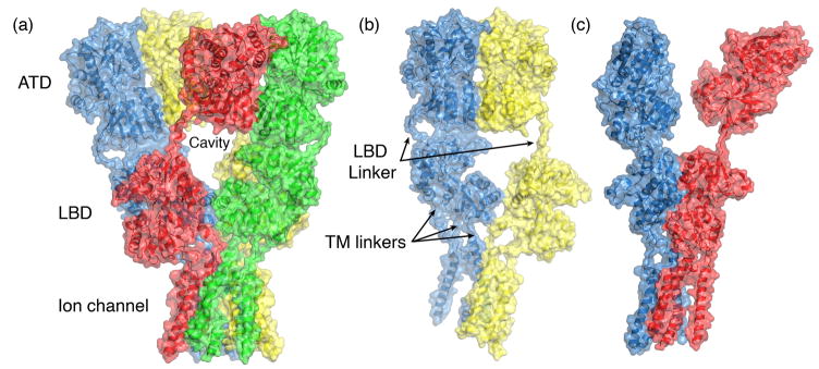
    1. Sobolevsky AI, Rosconi MP, Gouaux E. X-ray structure, symmetry and mechamism of an AMPA-subtype glutamate receptor. Nature. 2009;462:745–756. The 1st full length crystal structure of an iGluR tetramer, which took more than 5 years to solve, confirmed many predictions made from the structures of soluble iGluR domains, but also revealed many unexpected features which have no counterpart in other neurotransmitter receptors including symmetry mismatch, large cavities, and subunit cross over between domains. - PMC - PubMed
    1. Nakagawa T, Cheng Y, Ramm E, Sheng M, Walz T. Structure and different conformational states of native AMPA receptor complexes. Nature. 2005;433:545–549. - PubMed

Publication types

MeSH terms

Substances