. 2011 Apr;32(4):709-13.
doi: 10.3174/ajnr.A2366. Epub 2011 Feb 24.

Frequency and location of dilated Virchow-Robin spaces in elderly people: a population-based 3D MR imaging study

Affiliations

Frequency and location of dilated Virchow-Robin spaces in elderly people: a population-based 3D MR imaging study

Y-C Zhu et al. AJNR Am J Neuroradiol. 2011 Apr.

Abstract

Background and purpose: dVRS have been previously associated with aging and cerebrovascular diseases. However, little is known about their prevalence and topographic distribution in the general elderly population.

Materials and methods: dVRS were evaluated by using high-resolution 3D MR imaging in 1826 subjects enrolled in the 3C-Dijon MR imaging study. On T1-weighted MR imaging, dVRS were detected according to 3D imaging criteria and rated by using 4-level severity scores based in the BG or in the WM. The number and anatomic location of large dVRS (≥3 mm) were recorded.

Results: dVRS were observed in the BG or WM in every subject. The severity of dVRS was significantly associated with higher age in both the BG and WM, whereas sex was related to the severity of dVRS only in the BG. Large dVRS were detected in 33.2% of participants. Status cribrosum was found in 1.3% of participants. dVRS were also highly prevalent within the hippocampus (44.5%) and hypothalamus (11.6%).

Conclusions: dVRS are always detected in the BG or WM in elderly people, and large dVRS are also prevalent. The topographic distribution of dVRS is not uniform within the brain and may depend on anatomic or pathologic characteristics interacting with aging and sex.


Figures
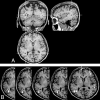
Fig 1.
Fig 1.
3D T1-weighted images from 1 participant showing multiple dVRS. A, Cystiform dVRS are analyzed on axial, coronal, and sagittal planes. B, Multiple dVRS exhibit a linear shape and orientation along cortical medullary arteries (thin white arrows) in the white matter. Cystic dVRS (large white arrows) are detected on contiguous sections showing a cavity with a vascular shape extension.
Fig 2.
Fig 2.
Locations of dVRS. A, In coronal sections, the upper white line crosses the superior limit of the caudate on both sides; the lower one crosses the base of the forebrain at the level of the anterior perforated substance. The lower one-third of the section between these 2 lines (between the black line and lower white line) is defined as belonging to the proximal part of the LSA, whereas the upper two-thirds (between the black line and upper white line) is considered as the distal part of the LSA. Large dVRS are detected in the proximal part of the LSA (white arrows) on both sides; a linear dilated Virchow-Robin space is also detected in the distal part of the LSA (black arrow). B, Large dVRS (diameter of 3 mm) are detected in the left distal part of the LSA (white arrows). C, Two Virchow-Robin spaces are seen in the left side of the hippocampus (arrows). D, A Virchow-Robin space is seen in the in right hypothalamus (arrows), as a small dot in the axial section and as a vascular shape in the longitudinal section.
Fig 3.
Fig 3.
Severity scores of dVRS in the BG.
Fig 4.
Fig 4.
Severity scores of dVRS in the WM.

References

    1. Maclullich AM, Wardlaw JM, Ferguson KJ, et al. . Enlarged perivascular spaces are associated with cognitive function in healthy elderly men. J Neurol Neurosurg Psychiatry 2004;75:1519–23 - PMC - PubMed
    1. Heier LA, Bauer CJ, Schwartz L, et al. . Large Virchow-Robin spaces: MR-clinical correlation. AJNR Am J Neuroradiol 1989;10:929–36 - PMC - PubMed
    1. Udaka F, Sawada H, Kameyama M. White matter lesions and dementia: MRI-pathological correlation. Ann N Y Acad Sci 2002;977:411–15 - PubMed
    1. Patankar TF, Mitra D, Varma A, et al. . Dilatation of the Virchow-Robin space is a sensitive indicator of cerebral microvascular disease: study in elderly patients with dementia. AJNR Am J Neuroradiol 2005;26:1512–20 - PMC - PubMed
    1. Wuerfel J, Haertle M, Waiczies H, et al. . Perivascular spaces: MRI marker of inflammatory activity in the brain? Brain 2008;131:2332–40 - PubMed

Publication types

MeSH terms